Laurie Anne Walden, DVM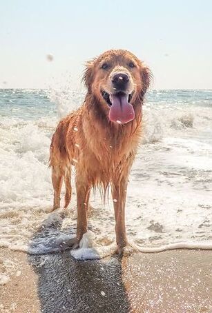 Photo by Elisa Kennemer on Unsplash Photo by Elisa Kennemer on Unsplash Warm weather brings some extra risks for pets. Some animals need protection from sunlight, and all animals need protection from hot temperatures.
Sun-Related Skin Conditions Ultraviolet (UV) radiation in sunlight causes skin damage in animals just like it does in humans. Fur and melanin (dark pigment) protect the skin from UV rays, so animals with short white hair or no hair are at higher risk than those with thick dark fur. Most animals with sun-related skin disease spend a lot of time outdoors, but even indoor cats can develop skin disease caused by UV rays coming through windows. Sun-related skin damage typically affects areas of the body that don’t have thick fur and are exposed to the sun. Any part of the body with thin or light-colored hair can be involved, but these are the most common areas:
Solar dermatitis, also called actinic dermatitis, is skin disease caused by exposure to UV radiation. It can be mistaken for allergic skin disease because the lesions are similar and it is often seasonal. Affected skin is red, painful to the touch, and scaly or flaky. Bumps and oozy lesions might develop. Over time, the skin becomes thickened and scarred. Because solar dermatitis usually causes a secondary skin infection, the lesions might improve with antibiotics, at least at first. UV radiation causes mutations within skin cells, so solar dermatitis can transform into skin cancer (squamous cell carcinoma or hemangiosarcoma, for example). These cancers are malignant: they can spread throughout the body. Solar dermatitis and skin cancer are diagnosed with skin biopsy. Heat Stroke Heat stroke is a life-threatening risk to animals during warm weather. Unlike solar dermatitis and skin cancer, it doesn’t require direct exposure to UV rays. The outdoor temperature doesn’t even have to be very hot for an animal to develop heat stroke. The temperature inside a parked car quickly rises higher than the outside temperature, so animals inside parked cars are at risk even in moderately warm weather. Brachycephalic (short-nosed) animals like pugs and bulldogs are at especially high risk. Signs of heat stroke include panting, dark red or purple gums, vomiting, collapse, and seizures. Heat stroke requires immediate first aid (cooling with water, not ice) and emergency veterinary care. For more information, see the blog post on heat stroke. How to Protect Your Pet The best way to protect animals is to minimize their exposure to UV rays and heat:
Sources
Image source: Elisa Kennemer on Unsplash Laurie Anne Walden, DVM Photo by Wall Boat on Flickr Photo by Wall Boat on Flickr Uveitis is inflammation inside the eye. Some of the many conditions that cause uveitis affect the whole body, so a diagnosis of uveitis often triggers a series of tests to find the cause.
The uvea is a pigmented layer within the eye. The iris, which gives the eye its color, is the part of the uvea that you can see when you look at your pet’s eyes. The other structures of the uvea (ciliary body and choroid) are behind the iris, sandwiched between other layers of the eye. The uvea has a blood supply to deliver nutrients to the eye, so anything that can travel through blood vessels—like infectious organisms and cancer cells—can reach the uvea and cause uveitis. The uvea also produces and drains fluid within the eye. Inflammation of the uvea interferes with fluid balance inside the eye, so untreated uveitis can lead to glaucoma (increased eye pressure) and blindness. Causes Some of the things that cause uveitis are problems with the eye itself:
Problems outside the eye can also cause uveitis:
Signs Uveitis causes the same nonspecific signs of eye trouble (like redness) as nearly every other eye disorder. It can also cause changes that are more specific to inflammation inside the eye, like a change in iris color. Animals with uveitis sometimes have no obvious signs at all. These are some of the signs of uveitis:
Diagnosis Uveitis is diagnosed by examining the eye with an ophthalmoscope to look for signs of inflammatory debris inside the eye. The eye exam might be the only diagnostic test needed if it reveals an eye problem that’s causing the uveitis. If the animal doesn’t have an obvious eye abnormality causing the inflammation, the next step is to conduct diagnostic tests to find the cause. The animal will most likely have baseline blood tests, tests for various infectious organisms (especially viruses and the many organisms carried by fleas and ticks), and possibly x-ray imaging or ultrasound. The patient might need to be referred to a veterinary ophthalmologist. Treatment Uveitis is treated with medication to reduce inflammation. This medication is usually in the form of eye drops or eye ointment. Some animals need oral medication in addition to eye medication. The purpose of this anti-inflammatory medication is to relieve pain and reduce the risk of glaucoma. Whatever caused the uveitis also needs to be treated. The prognosis for uveitis depends on the cause. If uveitis is diagnosed quickly and the underlying cause is treatable, the prognosis is generally good. Some conditions that cause uveitis have a more guarded prognosis. Image source: Wall Boat on Flickr Laurie Anne Walden, DVM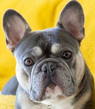 This French bulldog has narrowed nostrils and prominent facial skin folds. Photo by Dan Blackburn on Unsplash. This French bulldog has narrowed nostrils and prominent facial skin folds. Photo by Dan Blackburn on Unsplash. The French bulldog is now the most popular dog in the United States, according to the American Kennel Club (which defines popularity by the number of purebred dog registrations). This result is no surprise; Frenchies have been rising in the popularity ranks for several years. But these little dogs have a host of potential health problems, just like other animals bred to have flat faces and short noses.
Brachycephalic Head Shape Short-headed (brachycephalic) animals like English bulldogs, French bulldogs, pugs, and Persian cats have a shortened upper jaw and nose. The lower jaw is typically not shortened, so when they close their mouths, their lower incisor teeth might stick out in front of the upper incisors. Brachycephaly affects only the bones, not the soft tissues (skin, tongue, soft palate, and so forth), so brachycephalic animals have too much soft tissue for their face size. This is why brachycephalic animals have skin folds between their nose and eyes. They have the skin to cover a nose that just isn’t there. Respiratory Problems In a brachycephalic animal, the soft tissues inside the mouth and throat are crammed into an upper jaw that’s too short to hold them. All of this excess tissue blocks the airway, causing a cascade of problems related to the increased effort of breathing. The term brachycephalic airway syndrome describes problems caused by anatomic abnormalities that are common in brachycephalic animals. These abnormalities include narrowed nostrils, an elongated soft palate, and everted laryngeal saccules (tissue near the vocal cords that is pulled into the airway because of labored breathing over time). Brachycephalic animals might also have an enlarged tongue. Some have an abnormally narrow trachea, which increases the work of breathing and the risk of problems during anesthesia. Other Problems Brachycephalic animals are more likely than others to have heat stress. Their relatively shallow eye sockets and large eyelid openings increase their chance of developing corneal ulcers, dry eye, and other eye problems. Infections can develop between skin folds. Some brachycephalic animals have digestive tract problems like chronic vomiting, possibly related to gastric reflux caused by chronic labored breathing. They usually have crowded or malpositioned teeth. Natural birth is not possible for some brachycephalic dogs because the mom’s pelvis is too narrow for the puppies’ heads. Some breeds—including French bulldogs—usually require cesarean (surgical) delivery. Signs If you have a brachycephalic pet, watch for these signs of trouble:
Treatment Anatomic problems like narrowed nostrils, elongated soft palate, and everted laryngeal saccules can be corrected with surgery. You can see photos of these problems and more information about surgery on the American College of Veterinary Surgeons website: https://www.acvs.org/small-animal/brachycephalic-syndrome. One “treatment” for brachycephalic airway syndrome is to stop breeding dogs for extreme face shape. If you’re thinking of getting a French bulldog or another brachycephalic pet, reward breeders who breed for good health: choose a dog with round (not slit-like) nostrils, minimal or no skin folds near the eyes, and the ability to run, play, and sleep while breathing freely without snorting or gagging. Image source: Dan Blackburn on Unsplash Laurie Anne Walden, DVM Photo by Hermes Rivera on Unsplash Photo by Hermes Rivera on Unsplash Eyelid problems are common in dogs and cats. Some eyelid disorders cause damage to the eyeball itself and can affect vision. A pet with eye redness, eye drainage, eyelid swelling, an eyelid lump that is new or changing, or signs of eye discomfort (squinting, pawing at the eyes, rubbing the face) should see a veterinarian.
Benign Masses In dogs, most eyelid masses are benign tear gland tumors called meibomian gland adenomas. These masses are located along the lid margin and typically look like raised, bumpy, pink or dark brown lumps. Other types of benign tumors also occur in dogs and cats. A chalazion is a firm lump caused by a blocked tear gland. This type of lump is near the lid margin under the skin, not right on the lid margin like a meibomian gland adenoma. Benign eyelid masses can irritate the cornea (the clear structure at the front of the eye) and can enlarge over time. These masses are usually removed surgically. Removing an eyelid mass means removing part of the eyelid, so it’s best to do the procedure while the mass is still small. Cancerous Masses Most eyelid masses in cats, and some in dogs, are malignant cancers. Many types of cancer affect the eyelids, just like the rest of the skin; some examples are squamous cell carcinoma, melanoma, and mast cell tumor. The only way to know for sure if an eyelid lump is benign or malignant is to send it to a laboratory for analysis. If an eyelid mass is large or located near the inner corner of the eye, referral to a veterinary ophthalmologist is often the best option in case the patient needs reconstructive eyelid surgery. Cherry Eye The term cherry eye is an informal name for prolapse of the gland of the third eyelid. The third eyelid (nictitating membrane) is a pink structure at the inner corner of dogs’ and cats’ eyes. In animals with cherry eye, a tear gland protrudes out of its normal location behind the third eyelid; it looks like a smooth pink or red lump. Cherry eye is treated by surgically replacing the gland in its normal position. The prolapsed gland shouldn’t be removed entirely because it produces tears. Removal of the gland would put the animal at risk of dry eye (keratoconjunctivitis sicca). Swollen Eyelids Eyelid swelling could be caused by an allergic reaction, a benign or cancerous mass, inflammation, or infection. Eyelid swelling due to an allergic reaction is sudden and often affects both eyes. Because eyelid swelling is sometimes the first sign of a more serious reaction called anaphylaxis, it always warrants a call or visit to a veterinary clinic. An animal that has swollen eyelids along with other concerning symptoms like vomiting or wheezing should be taken immediately to an emergency clinic. Entropion (Rolled-in Eyelids) In animals with entropion, one or more eyelids are rolled inward so eyelashes and hair rub on the cornea. Entropion can be genetic or breed associated, especially in animals with loose facial skin and animals that are brachycephalic (short faced). The condition can also be caused by scarring, other eyelid disorders, or excessive squinting from eye discomfort. The damage to the cornea is uncomfortable at best and can eventually impair vision. Entropion is corrected with surgery. Young animals with entropion can sometimes be treated with a simple tacking procedure to help the eyelids roll back out to the normal position as they grow. Abnormal Hairs Occasionally hairs similar to eyelashes grow in the wrong location, either along the eyelid margin (distichia) or inside the eyelid (ectopic cilia). These hairs can cause significant pain and corneal ulcers. They’re hard to see and are typically found only with magnification during an ophthalmic examination to find out why an animal has a corneal ulcer. If they’re causing discomfort, the hairs and their follicles need to be removed. Eyelid Inflammation Inflamed eyelids are red and swollen, sometimes with oozy discharge and visible sores along the margins. The hair on the eyelid might be thin or missing. Eyelid inflammation has many possible causes, including immune-mediated disease, parasites (like mange mites), bacterial or fungal infections, and irritants. Diagnosis might require skin scrapings, cultures, and biopsy. Treatment depends on the underlying cause. More Information You can see photos of some of these conditions on the American College of Veterinary Ophthalmologists website: https://www.acvo.org/common-conditions1 Image source: Hermes Rivera on Unsplash Laurie Anne Walden, DVM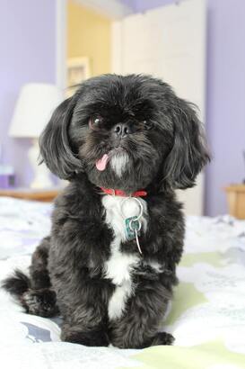 Photo by Jennifer Shishmanian on Unsplash Photo by Jennifer Shishmanian on Unsplash Keratoconjunctivitis sicca (KCS), or dry eye, is common in dogs and can also affect cats. KCS is caused by a problem with the quantity or quality of the tear film. The condition is uncomfortable and can cause blindness. For most patients, KCS is managed with lifelong eye medication.
Causes Tears are produced by glands in the eyelids and third eyelid. The tear film contains water, mucus, and oil. Deficiencies in any of the components of the tear film compromise its functions: lubricating the cornea (the clear structure at the front of the eye), removing debris, and providing nutrition to the cornea. Anything that affects the tear-producing glands can cause KCS. Brachycephalic (flat-faced) animals with protruding eyes that aren’t completely covered by the eyelids also develop KCS symptoms because of excessive water evaporation from the surface of the cornea. These are some of the causes of KCS:
Signs Animals with KCS typically have thick, sticky eye discharge. The discharge is thick because the watery part of the tear film is decreased or absent, so the tear film that remains is made mostly of mucus and oil. Either 1 or both eyes are affected, depending on the cause of the KCS. Tear film deficiencies increase the risk for corneal ulcers, long-term damage to the cornea, and impaired healing of the cornea. These are signs of KCS and the resulting corneal damage:
Diagnosis The tear film quantity is measured with the Schirmer tear test, in which a sterile paper strip with measurement markings is placed inside the lower eyelid for 1 minute. The distance that the tear film is absorbed along the strip shows whether the animal has low tear production. Veterinary ophthalmologists use other tests to find out whether a patient has a problem with tear film quality. Treatment Animals with KCS are treated with a combination of eye medications, most of which need to be applied 2 or 3 times a day. Medications include tear stimulants that reduce immune-mediated inflammation, tear replacements to lubricate the eye, and other medications to treat corneal damage. KCS can be managed but not cured, so medical treatment needs to continue for the rest of the animal’s life. For animals whose KCS doesn’t respond to eye medication, a few surgical options are available. The most common of these is parotid duct transposition, which involves rerouting a salivary duct from the mouth to the inside of the lower eyelid so the animal basically drools onto the eye. This procedure is performed by a veterinary ophthalmologist. Image source: Jennifer Shishmanian on Unsplash Laurie Anne Walden, DVM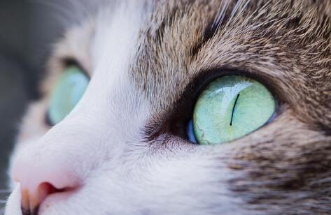 Photo by Antonio Lapa on Unsplash Photo by Antonio Lapa on Unsplash The conjunctiva is a thin membrane that covers the insides of the eyelids and the white part of the eye. Inflammation of the conjunctiva, or conjunctivitis, makes the eye look red. Conjunctivitis is sometimes called pinkeye. This name is a bit misleading, though, because lots of other eye disorders—some very serious—also cause redness of the eye.
The conjunctiva is a mucous membrane, like the membranes lining the nose and mouth. It protects the eye, helps lubricate the surface of the eye with tears, aids in eye movement, and is part of the system that heals damage to the cornea. Causes Conjunctivitis has many possible causes in dogs and cats:
Signs Signs include eye redness, squinting, blinking more than usual, and eye discharge. The discharge can be watery and clear or it can be cloudy, yellow, or greenish. The tissues around the white of the eye and the insides of the lids might look puffy. The animal might rub or paw at the eye because of itching or discomfort. Diagnosis Conjunctivitis can’t be diagnosed just by the appearance of the eye. Redness and discharge are part of the eye’s response to any abnormality, so other problems need to be ruled out before conjunctivitis can be diagnosed. Diagnosis includes a general physical examination, an eye examination with an ophthalmoscope, and a corneal stain to identify ulcers or other damage to the cornea. Other tests include measuring tear production, measuring eye pressure (to rule out glaucoma), and sometimes swabbing or scraping the conjunctiva to obtain samples for laboratory analysis. Treatment Conjunctivitis is treated with topical medication (eye drops or eye ointment); oral medication is sometimes also used. The type of medication depends on the cause of the conjunctivitis. Antibiotics are usually part of the treatment, either to eliminate a primary bacterial infection or to prevent a secondary bacterial infection. Cats with known or suspected herpesvirus conjunctivitis sometimes need antiviral medication. Eye medications used to treat conjunctivitis often contain a steroid (such as dexamethasone or hydrocortisone) to reduce inflammation. However, steroids interfere with corneal healing, so before a steroid-containing medication is used, the cornea must be stained and examined with an ophthalmoscope to be sure there is no corneal ulcer or scratch. If your pet has eye medication from an earlier episode of conjunctivitis, don’t use it to treat a new episode of eye redness unless your veterinarian has instructed you to. And never use any over-the-counter (nonprescription) eye product in an animal without first consulting your veterinarian. Photo by Antonio Lapa on Unsplash Laurie Anne Walden, DVM Public domain photo from pixy.org Public domain photo from pixy.org Corneal ulcers are common causes of eye redness in dogs and cats. A corneal ulcer is an area of damage to the surface of the cornea, the clear structure at the front of the eye. These ulcers are painful but usually heal readily with treatment. Sometimes corneal ulcers develop complications that require surgery or other interventions.
Causes Anything that scratches, hits, or irritates the surface of the eye can cause a corneal ulcer. Animals with low tear production are at increased risk for corneal ulcers because they have a low volume of tears to wash away irritants. These are some of the things that cause corneal ulcers:
Complications Some animals have a defect of the corneal epithelium—the surface layer of corneal cells—that increases the risk of ulcers and keeps ulcers from healing. These slow-healing ulcers are called indolent ulcers. Corneal ulcers can become infected, threatening the animal’s vision. Deep corneal ulcers and wounds can penetrate all the way through the cornea and rupture the eye. Eye medications that contain steroids (commonly used to treat conjunctivitis and other inflammatory eye conditions) prevent corneal ulcers from healing properly. Steroid-containing eye medications can also increase the risk of corneal infection. Signs The signs of eye discomfort are the same whether they are caused by a corneal ulcer or by another eye disorder. Typical signs are squinting, redness of the white part of the eye, and clear or cloudy eye drainage. The animal might rub the eye, and the cornea might look cloudy. Deep ulcers are sometimes visible without an ophthalmoscope and look like a dent or divot in the cornea. Superficial ulcers are usually not visible to the naked eye. An animal with any of these signs should see a veterinarian right away. Although uncomplicated corneal ulcers are relatively minor, they are painful. Some of the conditions that cause the same signs are medical emergencies that require immediate treatment to save the eye. Diagnosis Corneal ulcers are diagnosed with fluorescein dye applied to the eye. This green dye shows areas where the normal corneal epithelium is disrupted or missing. The eye is also examined under magnification to assess the depth and size of the ulcer, detect complications, and find the cause. Treatment An uncomplicated ulcer is treated with eye medication to prevent infection and relieve pain. The animal might be fitted with a protective collar (the lampshade type) to prevent eye rubbing. The cause of the ulcer is also treated, if possible. The eye is rechecked with fluorescein dye after a few days to be sure the ulcer has fully healed. Complicated ulcers are treated according to their cause and severity. Indolent ulcers are treated with a procedure to remove loose corneal epithelium. This procedure can often be performed in an awake animal with drops to numb the eye. Some indolent ulcers need a more extensive procedure with the animal under general anesthesia. Deep ulcers and wounds that might rupture the eye need surgical treatment by a veterinary ophthalmologist. Image source: https://pixy.org/6427021/ Laurie Anne Walden, DVM Public domain image from pixy.org Public domain image from pixy.org Osteoarthritis is very common in cats but often goes undetected. In most cats, joint pain doesn’t cause obvious signs like limping. Instead, it causes changes in mobility and behavior that can be misinterpreted as normal aging. Cat owners’ recognition of these changes is the first step in diagnosing and relieving joint pain.
Cause Osteoarthritis is a chronic disease in which the protective cartilage in a joint wears down. Eventually the bones and other structures in the joint deteriorate, causing pain that worsens over time. Degenerative joint diseases like osteoarthritis can be caused by trauma or by problems with the structure of a joint, but often the cause is not known. Senior cats are by far the most likely to develop osteoarthritis. Younger cats can be affected too. Osteoarthritis in cats most often involves the hips, elbows, knees, and hocks. Cats can also develop degenerative joint disease in the spine. Various studies have shown that between about 60% and 90% of cats have evidence of degenerative joint disease on radiographs (x-ray images).[1-3] Not all of these cats have pain, though, at least not at first. Signs In cats, signs of joint pain are subtle. Cats tend to hide signs of pain. Osteoarthritis often affects joints on both sides of cats’ bodies, so they don’t develop lameness—it’s hard to limp with both front legs or both rear legs at the same time. Signs of osteoarthritis in cats reflect their limited mobility, reduced activities of daily living, and general grumpiness caused by chronic pain:
Diagnosis The most important diagnostic tool is cat owners’ observations of signs of joint pain at home. Treatment is often started just on the basis of behavior changes consistent with pain. Cat owners can use questionnaires like the Feline Musculoskeletal Pain Index (https://painfreecats.org/) to record and score their cats’ signs of pain. These assessment tools help veterinarians diagnose joint pain and are also very useful to track changes over time and monitor response to treatment. Cats with signs of pain should receive a physical examination to be sure the signs are caused by joint or back pain and not by something else. A full orthopedic examination of a cat is challenging (sort of like examining an uncooperative bowl of jello that doesn’t tell you when it hurts and won’t trot on leash), but sometimes an examination reveals joint thickening or other physical changes of osteoarthritis. Radiographs can show evidence of degenerative joint disease but aren’t always needed. The decision to use imaging depends on the individual cat. Treatment Never give a cat pain medication, including nonprescription over-the-counter remedies, without consulting a veterinarian. Some medications that are safe for people and dogs are very dangerous for cats. In cats, joint pain is managed with a combination of nondrug and drug treatments. A multimodal approach (using several strategies) tailored to each cat’s pain level and living conditions is the best way to help relieve chronic pain in cats. Nondrug treatments include weight management, adjunctive therapies like acupuncture, dietary supplements such as glucosamine, and environmental modifications like ramps, steps, soft bedding, and litter boxes with low sides. Drug options are more limited for cats than they are for dogs, but a number of drugs are available. A new injectable treatment for osteoarthritis pain avoids the need to give a cat a pill by mouth.[3] References 1. Lascelles BD, Henry JB 3rd, Brown J, et al. Cross-sectional study of the prevalence of radiographic degenerative joint disease in domesticated cats. Vet Surg. 2010;39(5):535-544. doi:10.1111/j.1532-950X.2010.00708.x 2. Slingerland LI, Hazewinkel HA, Meij BP, Picavet P, Voorhout G. Cross-sectional study of the prevalence and clinical features of osteoarthritis in 100 cats. Vet J. 2011;187(3):304-309. doi:10.1016/j.tvjl.2009.12.014 3. Gruen ME, Myers JAE, Lascelles BDX. Efficacy and safety of an anti-nerve growth factor antibody (frunevetmab) for the treatment of degenerative joint disease-associated chronic pain in cats: a multisite pilot field study. Front Vet Sci. 2021;8:610028. doi:10.3389/fvets.2021.610028 4. Bennett D, Zainal Ariffin SM, Johnston P. Osteoarthritis in the cat: 1. How common is it and how easy to recognise? J Feline Med Surg. 2012;14(1):65-75. doi:10.1177/1098612X11432828 Image source: https://pixy.org/6310117 Laurie Anne Walden, DVM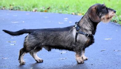 Intervertebral disk disease (IVDD), also referred to as slipped disk, ruptured disk, or herniated disk, is a relatively common cause of neck and back pain in dogs. Some dogs with IVDD also develop neurologic deficits like toes knuckled under, a wobbly gait, or the inability to walk. The prognosis depends on the severity of the injury and length of time before treatment. A sudden inability to walk is a medical emergency. Affected dogs might need urgent surgery.
Anatomy The spine is a row of individual bones called vertebrae. The spinal cord runs along a bony canal through the vertebrae. Intervertebral disks sit between the vertebrae just below the spinal cord and provide cushioning and support for the spine. Intervertebral disks are sort of like jelly doughnuts: they have a squishy interior (the nucleus pulposus) and a firmer, protective exterior (the annulus fibrosus). With IVDD, part of a disk herniates; it leaks or bulges beyond its normal location and presses on the spinal cord or on the surrounding nerves. Types of IVDD IVDD is classified according to the type of disk herniation. Type 1 IVDD has a genetic link: it’s most common in dog breeds with chondrodystrophy, which usually means dogs with short legs and a long back. In type 1 IVDD, the nucleus pulposus becomes mineralized (hardens) over time and eventually breaks through the annulus fibrosus into the spinal canal, bruising the spinal cord. This type of herniation can happen quickly, causing sudden symptoms. Affected dogs are often young adults. Type 2 IVDD is caused by age-related breakdown of the annulus fibrosus. Weakening of the annulus fibrosus allows the disk to bulge upward against the spinal cord. Type 2 IVDD is more gradual than type 1 and is more common in older dogs. Intervertebral disk herniation can also be caused by trauma or strenuous exercise. Dogs at Risk Any dog can develop IVDD. These are the breeds at highest risk for type 1 IVDD:
Type 2 IVDD is more common in larger dogs like these:
Symptoms The symptoms of IVDD range from pain to paralysis, depending on the severity of herniation, the force with which the herniated disk material hits the spinal cord, and the duration of the injury. The mildest form of IVDD is neck or back pain with ability to walk and no neurologic deficits. Neck pain can cause limping, so the symptoms of a herniated disk in the neck can be mistaken for an orthopedic problem. More severe spinal injuries progress through stages that worsen with time: limb weakness, neurologic deficits, inability to walk, inability to stand, inability to move the limbs, and finally inability to feel pain in the limbs. Dogs with spine injuries might lose their normal bladder and bowel function. These are some of the symptoms of IVDD:
Diagnosis A physical examination shows whether the patient has neurologic deficits and often reveals the approximate location of the injury in the spine. Advanced imaging techniques like computed tomography or magnetic resonance imaging are required to make a definite diagnosis and locate all of the affected disks. Radiographs (x-ray images) are less helpful for diagnosing IVDD but might be obtained to rule out other possible causes of the symptoms, like fractures, dislocations, bone infection, and cancer. Treatment Dogs with milder symptoms—pain, ability to walk, and no or mild neurologic deficits—are usually treated medically at first. A crucial part of medical management is restricting the dog’s activity to allow the annulus fibrosus to heal, similar to treating a sprain. Some dogs need strict crate confinement. Medical management also includes medication to relieve pain. Dogs that begin to feel better with pain medication can reinjure themselves if they resume normal activity too soon, so they usually need to continue strict activity restriction for weeks. Dogs with neurologic deficits or pain that doesn’t improve with medical management often need surgery to remove herniated disk material and relieve pressure on the spinal cord. Time is of the essence; the longer the injury to the spinal cord is present, the worse the neurologic deficits and the worse the prognosis for recovery. For dogs that can’t walk but can still feel sensation in their feet, surgery performed as soon as possible after symptoms begin typically gives the best chance of being able to walk again. Once pain sensation in the feet is lost, the odds of recovery are much lower even with surgery. Spine surgery is expensive and dogs need extensive physical rehabilitation afterward, so the decision to proceed with surgery depends partly on the dog owner’s financial situation and ability to provide long-term care. Sources Olby NJ, Moore SA, Brisson B, et al. ACVIM consensus statement on diagnosis and management of acute canine thoracolumbar intervertebral disc extrusion. J Vet Intern Med. 2022. doi:10.1111/jvim.16480 Spinella G, Bettella P, Riccio B, Okonji S. Overview of the current literature on the most common neurological diseases in dogs with a particular focus on rehabilitation. Vet Sci. 2022;9(8):429. doi:10.3390/vetsci9080429 Photo by PublicDomainPictures on Pixabay Laurie Anne Walden, DVM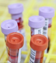 Photo by Daniel Sone, National Cancer Institute. Photo by Daniel Sone, National Cancer Institute. Laboratory testing of blood, urine, and stool samples is routinely done for animals. Abnormalities often show up on lab tests before an animal has obvious symptoms. Even healthy pets need to be tested regularly for parasites. Well-animal tests can detect problems early and help veterinarians track changes over time. These are some of the reasons your veterinarian might recommend lab tests for your pet:
Complete Blood Count A complete blood count measures the number, size, and shape of each type of cell in the blood.
Blood Chemistry Panel Blood chemistry tests measure substances in the blood that indicate changes in organ function or other biological processes. Blood chemistry analysis is run as a panel of many individual tests. A chemistry panel doesn’t always give a diagnosis for a sick animal, but the results can help the veterinarian narrow down the list of possibilities. Veterinary laboratories offer many chemistry panels for different species and diagnostic needs. A small panel that’s sufficient for a young animal before routine surgery might not be appropriate for an ill animal or an older pet. These are a few of the tests commonly included in chemistry panels:
Urinalysis Analysis of the urine includes specific gravity (a measure of urine concentration), pH, chemistry results like glucose and protein levels, and microscopic evaluation for cells, crystals, and bacteria. Urinalysis can reveal urinary tract infection, support a diagnosis of diabetes, and help evaluate the function of the kidneys and other organs. A complete laboratory analysis—especially for a senior pet—should include urinalysis. Parasite Tests Parasites are very common in pet animals (unless they have received regular parasite prevention as recommended by a veterinarian). Many of the parasites that pets carry are contagious to people. At least once a year, pets should have a stool test for intestinal parasites like hookworms and roundworms. Dogs should also have a yearly blood test for heartworm disease. Public domain image source: National Cancer Institute, Daniel Sone (photographer) |
AuthorLaurie Anne Walden, DVM Categories
All
Archives
February 2024
The contents of this blog are for information only and should not substitute for advice from a veterinarian who has examined the animal. All blog content is copyrighted by Mallard Creek Animal Hospital and may not be copied, reproduced, transmitted, or distributed without permission.
|
 RSS Feed
|
Office Hours
Monday through Friday 7:30 am to 6:00 pm
|
|


Site powered by Weebly. Managed by IDEXX Laboratories