Laurie Anne Walden, DVM Photo by Kari Shea on Unsplash Photo by Kari Shea on Unsplash Arterial thromboembolism (ATE) in cats has a guarded to poor prognosis and a high mortality/euthanasia rate. In ATE, a blood clot forms in the heart, travels through the circulation, and lodges in an artery. The clot blocks blood flow through that artery, causing significant pain and tissue damage. When the clot blocks an artery supplying a limb, as is usually the case, the limb becomes weak or paralyzed.
The most common site for ATE in cats is the aorta at the point where it divides into smaller arteries that supply the rear legs. A clot stuck here takes on the shape of a saddle, so ATE in this location is called a saddle thrombus. Cats with a saddle thrombus have sudden extreme pain and paralysis of the rear legs (usually both rear legs but sometimes just 1). Witnessing their pain and distress can be quite upsetting to their caretakers. Cause ATE is usually caused by heart disease. Unfortunately, a saddle thrombus can be the very first sign that a cat has heart trouble. The most common type of heart disease in cats—and thus the most common cause of ATE—is hypertrophic cardiomyopathy, an abnormal thickening of the heart muscle. Hypertrophic cardiomyopathy is most common in senior cats but can also affect younger cats. An estimated 15% of all cats and up to 29% of senior cats have hypertrophic cardiomyopathy.[1] Of these cats, a reported 11.3% develop ATE within 10 years after diagnosis.[2] Signs Signs of ATE usually appear suddenly. A cat with ATE will cry loudly in pain and might vomit. The cat will drag the affected limb or be unable to walk. The affected limb might feel cool to the touch. If the cat’s underlying heart disease has progressed to heart failure, the cat might also have signs like panting or rapid breathing. Diagnosis ATE can usually be diagnosed with a physical examination. The affected limb is typically cool, the nail beds are bluer or paler than those of the other paws, and the pulse in that limb is absent or weak. A heart murmur might be present. Diagnostic tests can be used to rule out other possible causes, especially if the cat has less obvious signs caused by a partial blockage. Treatment Many cats with ATE are euthanized before treatment is attempted. Factors linked to a better prognosis include involvement of only 1 limb, ability to move the affected limb, and absence of heart failure (meaning that the heart disease has not yet progressed to the level of heart failure). For cats that receive treatment, the immediate priority is pain control. They also receive anticoagulant medications (blood thinners), treatment for shock and tissue damage, and treatment for heart disease. Cats that don’t have heart failure and survive the first few days in the hospital have a chance of regaining the use of the affected limb once the clot dissolves. Prevention For cats known to have heart disease, certain findings on echocardiography (heart ultrasound) can indicate an increased risk for ATE. These cats can receive preventive anticoagulant medications. However, many cats with heart disease have no obvious signs, so their heart disease isn’t diagnosed. References 1. Luis Fuentes V, Abbott J, Chetboul V, et al. ACVIM consensus statement guidelines for the classification, diagnosis, and management of cardiomyopathies in cats. J Vet Intern Med. 2020;34(3):1062-1077. doi:10.1111/jvim.15745 2. Lo ST, Walker AL, Georges CJ, Li RH, Stern JA. Dual therapy with clopidogrel and rivaroxaban in cats with thromboembolic disease. J Feline Med Surg. 2022;24(4):277-283. doi:10.1177/1098612X211013736 Image source: https://unsplash.com/photos/gray-tabby-cat-FilM6ng7VGQ Laurie Anne Walden, DVM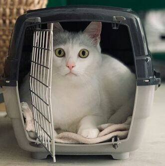 Photo by Anatolii Kozhukhar on Unsplash Photo by Anatolii Kozhukhar on Unsplash Many dogs and cats experience fear, anxiety, and stress at the veterinary clinic. Their anxiety often begins before they even arrive at the clinic, especially if they associate pet carriers and car rides with the clinic. Previsit antianxiety medications for pets can make clinic visits easier and safer for these pets, their owners, and clinic staff.
These are some of the benefits of previsit medications for anxious pets:
Previsit medications have benefits for pet owners and clinic staff members too. Being the caretaker of an anxious and fearful pet isn’t easy, so previsit medications for pets can reduce everyone’s stress levels. For clinic staff members, managing patients’ clinic anxiety reduces the risk of injury and decreases workplace stress (we love animals too and don’t want to cause them distress). How to Know if Your Pet Needs Previsit Medications Previsit medications aren’t just for aggressive animals. Pets with more subtle signs of stress are scared too, and they deserve help for their anxiety just as much as animals that can’t be handled without full sedation. If your pet shows obvious signs of fear at the clinic, you probably already know that your pet would benefit from previsit medications. But maybe your pet’s signs are not as noticeable and you’re a little surprised when your veterinarian recommends medications. Dogs and cats show anxiety and fear with a range of behaviors and body language cues. Anxious and fearful dogs and cats tend to start with subtle behaviors like lip licking or freezing. If the scary thing doesn’t go away—or tries to draw a blood sample—the behavior can escalate to aggression. Dogs and cats that are anxious at the veterinary clinic can show these signs:
How to Use Previsit Medications Previsit medications work best if they’re used along with training and positive reinforcement. For example, a cat can make positive associations with the cat carrier if the carrier is left out all the time as part of the normal household furniture, sometimes with yummy treats inside, instead of appearing only when the cat is going to the clinic. Veterinarians choose previsit medications according to the needs and medical condition of the individual patient. Some of the drugs commonly prescribed for situational (short-term) anxiety are gabapentin, trazodone, and clonidine. Motion sickness contributes to anxiety, so pets might also receive antinausea medication. Some of these medications can cause sedation, but the main goal is anxiety reduction, not sedation. Previsit medications are timed to have maximum effect at the time of the clinic visit. They need to take effect before the pet experiences any stress. Most of these medications are given at least 1 to 2 hours before the clinic visit; sometimes a loading dose is also given the night before. They typically last for up to 8 hours, although the duration can vary. Because individual animals respond differently to these medications, giving a trial dose can be very helpful. If you use a trial dose, tell your veterinarian how long it took for the medication to take effect, how the medication affected your pet’s behavior, whether your pet had adverse effects like vomiting or excessive sedation, and how long the medication’s effects lasted. For some dogs and cats, previsit medications aren’t enough to overcome their fear. These pets might also need a sedative injection at the clinic. For these pets, previsit medications given at home make the sedative injection easier for the pet (and staff) and can also reduce the dose of injectable drug. Image source: https://unsplash.com/photos/a-white-cat-sitting-inside-of-a-cage-B-_SyRxv2So Laurie Anne Walden, DVM Photo by Hal Gatewood on Unsplash Photo by Hal Gatewood on Unsplash Over-the-counter (nonprescription) pain medications can cause serious problems for dogs and cats. Because these medications don’t need a prescription and are used for children as well as adults, some pet owners mistakenly believe that they’re safe for animals too. But dogs and cats don’t process these drugs the same way as humans.
If your pet has signs of pain, call your veterinarian instead of giving your pet something from your medicine cabinet. Prescription pain medications developed specifically for dogs and cats are safer and more effective for them than over-the-counter human medications. In any case, animals that have a level of pain high enough to be obvious to a human need to see a veterinarian. Nonsteroidal Anti-Inflammatory Drugs (Aspirin, Ibuprofen, Naproxen) The most common human nonsteroidal anti-inflammatory drugs (NSAIDs) available without a prescription are aspirin, ibuprofen, and naproxen. Some brand names are Advil, Aleve, Ascriptin, Bayer, Bufferin, Ecotrin, Midol, and Motrin. In animals, NSAIDs can cause these problems:
NSAIDs reduce inflammation by blocking the activity of cyclooxygenase (COX) enzymes, which are responsible for prostaglandin production. Prostaglandins are hormone-like substances with many functions; they’re involved in the inflammatory response and also protect the stomach lining and maintain blood flow to the kidneys. Because NSAIDs decrease prostaglandin levels, these medications decrease inflammation and pain. However, the lower prostaglandin levels can also cause serious adverse effects like stomach ulcers and kidney damage. Some newer NSAIDs target specific COX enzymes and prostaglandins that are less likely to affect the stomach and kidneys. Several NSAIDs have been approved for use in dogs, and a few are available for cats. Examples are carprofen, grapiprant, deracoxib, and robenacoxib. These species-specific drugs are available only by prescription from a veterinarian and are less likely than human NSAIDs to cause adverse effects in dogs and cats. Animals receiving prescription NSAIDs need regular examinations and bloodwork to monitor kidney and liver function. Acetaminophen Brands containing acetaminophen include Tylenol, Panadol, Excedrin, and Midol. Some pain relievers labeled “complete” or “dual action” contain acetaminophen plus aspirin or ibuprofen. Acetaminophen is also called paracetamol. Acetaminophen is highly toxic and often fatal to cats. Acetaminophen is also potentially toxic to dogs, but it can be used in dogs with caution and veterinary oversight. In humans and dogs, acetaminophen is broken down mainly in the liver through a process called glucuronidation. A toxic dose of acetaminophen in a human or a dog damages liver cells and causes liver failure. Cats don’t have the enzymes needed for glucuronidation, so their bodies break down acetaminophen through a different process called sulfation. The sulfation process in cats creates products that react to hemoglobin, the substance within red blood cells that carries oxygen throughout the body. In cats, acetaminophen exposure causes anemia and life-threatening oxygen deficiency. Cats that swallow acetaminophen are less likely than dogs to develop liver failure because they usually die of oxygen deficiency before the liver has a chance to fail. An antidote to acetaminophen (N-acetylcysteine) is available, so dogs and even cats with acetaminophen poisoning can recover if they are treated immediately at an emergency veterinary clinic. For More Information Get the Facts About Pain Relievers for Pets (US Food and Drug Administration): https://www.fda.gov/animal-veterinary/animal-health-literacy/get-facts-about-pain-relievers-pets Image source: https://unsplash.com/photos/round-white-pills-iPl3q-gEGzY Laurie Anne Walden, DVM Photo by Anya Prygunova on Unsplash Photo by Anya Prygunova on Unsplash In recent years, monoclonal antibodies have been approved to treat some common medical conditions in animals:
More monoclonal antibodies are likely to become available for dogs and cats. Monoclonal antibodies are used in humans to treat infections, autoimmune disorders, cancer, and other conditions, and they’re being investigated for similar uses in dogs and cats. How Monoclonal Antibodies Work Monoclonal antibodies are in a therapy class called biologics, meaning therapies made from living sources like animal cells, plant cells, or microorganisms. Biologics are large molecules that interact with receptors on the outer surfaces of cells; they don’t enter cells. In contrast, most traditional drugs are small molecules that work inside cells. Monoclonal antibodies are similar to the natural antibodies produced by the immune system. When the immune system detects an antigen, different immune cells release lots of different antibodies that tag different sites on that antigen. (An antigen is anything that triggers an immune response.) Once the antibodies have tagged the antigen, other components of the immune system arrive to destroy it. Monoclonal antibodies are cloned in a laboratory from just one antibody made by one type of immune cell (mono means “one”). They tag only a single site on an antigen. Monoclonal antibody therapy can be very precise because it directs the immune response to a narrow target. In addition to tagging invaders like viruses, monoclonal antibodies can be engineered to tag a receptor on an animal’s own cells. The monoclonal antibodies that relieve pain in dogs and cats, for example, block production of a protein that’s partly responsible for the pain sensation. After monoclonal antibodies have activated the immune system, they’re broken down in the body the same way that natural antibodies are. Unlike small-molecule drugs that enter an animal’s cells, they aren’t cleared through the liver or kidneys. Administration Monoclonal antibodies are given by injection at a veterinary clinic. They can’t be given by mouth because they’re proteins and would be destroyed by digestion. Their effect lasts a few weeks (4-8 weeks for Cytopoint, 4 weeks for Solensia and Librela). These therapies are species specific: monoclonal antibodies for dogs work only for dogs, not for cats. Benefits The monoclonal antibodies that are available for dogs and cats generally work well for their approved uses. They are less likely than some traditional drugs to cause adverse effects because they have a narrow target of action and don’t enter cells. Because they are not cleared through the liver or kidneys, they can be safer than other drugs for animals with liver or kidney disease. Injections given every few weeks might be more convenient for pet owners than pills given multiple times a day, especially if their pets resist oral medication. Adverse Effects Monoclonal antibodies have fewer adverse effects than many traditional drugs. An antibody injection can cause an allergic reaction, although this is rare. Manufacturers might recommend precautions for individual monoclonal antibody products. Image source: https://unsplash.com/photos/a-dog-wearing-a-baseball-glove-E3K7xyAvQ8M Laurie Anne Walden, DVM Photo by Kanashi on Unsplash Photo by Kanashi on Unsplash Vestibular disorders cause problems with balance and coordination. Animals with vestibular disorders typically have a head tilt along with stumbling, walking in circles, and possibly vomiting.
Vestibular problems that start suddenly can look scary to pet owners. The signs can be mistaken for stroke. Some conditions that cause vestibular problems are serious, even life-threatening; others are relatively minor. One of the most common vestibular disorders in dogs is a benign condition called idiopathic vestibular syndrome (idiopathic means “of unknown cause”). This condition is not serious and gets better on its own with time. Idiopathic vestibular syndrome most often affects senior dogs, so in dogs it’s also known as old dog vestibular disease. Cats can also develop idiopathic vestibular syndrome. Causes The vestibular system includes the brainstem, cerebellum (part of the brain), inner ear, and nerves that conduct signals between the inner ear and brain. Anything that affects any part of this system can cause vestibular signs. Vestibular disorders are either central (related to the brainstem and cerebellum) or peripheral (related to the inner ear and surrounding nerves). Central vestibular disorders are usually much more worrisome than peripheral vestibular disorders. Idiopathic vestibular syndrome is a peripheral vestibular disorder. The exact cause in dogs isn’t known. Similar conditions in humans are caused by inflammation of the inner ear or surrounding nerves, viral infections, and benign positional vertigo (an inner ear problem). These same causes are possible but not proven in dogs. Some of the other causes of vestibular disease in animals are deep ear infections (common), cancer, drug adverse effects, toxins, systemic disorders like hypothyroidism, meningitis, and other neurologic disorders. Signs These are the typical signs of a vestibular disorder:
Idiopathic vestibular syndrome usually affects older dogs and cats. It begins suddenly and then starts to improve within a few days. The signs are most pronounced on the first day and don’t get worse with time. In most animals, the signs disappear after a couple of weeks, although the head tilt can last much longer. Other diseases that affect the vestibular system cause signs of their own. For example, a central vestibular disorder might cause seizures or changes in consciousness. Vestibular signs that get worse with time are a signal that the animal has something other than idiopathic vestibular syndrome. Diagnosis Idiopathic vestibular syndrome is a diagnosis of exclusion: it’s diagnosed after other possibilities are ruled out. Diagnosis begins with a neurologic examination, eye and ear examinations, baseline bloodwork, and usually a thyroid function test and urinalysis. Depending on the results, the animal might need referral to a veterinary neurologist and advanced tests like magnetic resonance imaging of the head or cerebrospinal fluid analysis. Treatment For idiopathic vestibular syndrome, the goal of treatment is to make the patient comfortable until the condition resolves on its own. Most animals receive prescription antinausea medication; some also need appetite stimulants or fluid therapy. Home care includes making sure they can eat and drink and protecting them from falling down steps or bumping into things like furniture corners. Physical therapy and postural maneuvers that are used to help people with vertigo aren’t commonly used in animals. Prognosis Most animals with idiopathic vestibular syndrome recover completely, although some have a permanent head tilt afterward. Some animals have more vestibular episodes later on. For vestibular signs that aren’t caused by idiopathic vestibular syndrome, the prognosis depends on whether the cause is treatable. Sources
Image source: https://unsplash.com/photos/brown-and-black-german-shepherd-Xy7SLX9zuVM Laurie Anne Walden, DVM Public domain photo by Ian Livesey on Flickr Public domain photo by Ian Livesey on Flickr Inflammatory bowel disease (IBD) causes chronic inflammation and irritation of the digestive tract. This condition is one of the most common chronic intestinal disorders in cats. IBD in cats is similar to Crohn disease in humans.
Cause In IBD, inflammatory cells accumulate within the walls of the digestive tract. The small intestine is the most common location in cats; the large intestine and stomach can also be affected. The intestinal walls become thickened and can’t absorb nutrients normally. Some cats with IBD also have inflammation of the pancreas and liver, a combination called triaditis. The cause of IBD isn’t known. The disease is probably caused by abnormal immune system function related to intestinal bacteria (which are necessary for normal digestion and immune function), diet, genetics, or other factors. IBD usually affects middle-aged or older cats, although cats of any age can develop the disease. Signs The signs are similar to those of many other digestive system disorders:
Diagnosis Because so many conditions cause the same signs as IBD, cats with chronic digestive tract problems need a thorough workup. Diagnostic tests are performed to rule out a long list of other possibilities, including intestinal lymphoma (a cancer that is clinically very similar to IBD), parasites, intestinal foreign bodies, bacterial and viral infections, food allergies, other types of cancer, and diseases like hyperthyroidism that cause general illness. Baseline tests include fecal parasite tests, bloodwork, and urinalysis. Additional blood tests are used to assess the function of the pancreas and check for evidence that the small intestine is not absorbing nutrients properly. Imaging (radiography and ultrasonography) is used to evaluate the digestive tract, liver, pancreas, and other organs. The only way to definitely diagnose IBD and distinguish it from intestinal lymphoma is to obtain biopsy samples from the intestine and submit them to a diagnostic laboratory for analysis. Biopsy is performed either with endoscopy (tube inserted through the mouth or anus) or abdominal surgery. Both of these procedures require general anesthesia. Treatment IBD is managed, not cured. Treatment is tailored to the individual cat, depending on what works best for that cat. Most cats receive a dewormer to eliminate parasites as a possible cause. Some cats’ signs improve with a hypoallergenic diet, so treatment can include several weeks of a food trial with a prescription diet. Antibiotics are sometimes used carefully (they can disrupt the normal intestinal bacteria). Probiotics and prebiotics, which contain beneficial bacteria or nutrients for existing intestinal bacteria, might be included in the treatment plan. Drugs that suppress the immune response, such as the corticosteroid prednisolone, are necessary for some cats. Prognosis Cats whose disease responds well to treatment can have a good quality of life, although their IBD signs will probably still flare up occasionally. The treatment type and medication doses often need to be adjusted over time. Image source: https://www.flickr.com/photos/ianlivesey/43270035161/ Laurie Anne Walden, DVM Photo by Anna Shvets on Pexels Photo by Anna Shvets on Pexels Home dental care is a crucial part of keeping dogs’ and cats’ mouths healthy. But many pet owners are reluctant to brush their pets' teeth (and brushing is not safe with animals that might bite), so the market is full of products that claim to clean teeth or freshen breath without brushing.
Some home dental care products work pretty well, some don’t work at all, and a few pose safety risks for pets. Here’s how to choose dental products for your pet. The Point of Home Dental Care The goal of cleaning your pet’s teeth at home is to remove plaque. Plaque, a sticky biofilm that contains millions of bacteria, attaches to tooth surfaces. Dental disease in dogs and cats is caused by plaque under and at the edges of the gums, not by tartar (hardened plaque) that you can see on tooth surfaces. Like humans, pets need a combination of professional dental cleanings and home dental care. Home dental products can’t remove tartar or treat dental conditions like periodontal disease. Some products make gum inflammation worse. Tartar and dental diseases require a dental procedure with general anesthesia at a veterinary clinic. When to Use Dental Products If your pet has smelly breath, visible tartar, bleeding gums, loose teeth, missing teeth, or signs of mouth pain, this is not the time to go shopping for dental products. This is the time to take your pet to the veterinarian and then schedule a dental procedure if your veterinarian recommends it. The time to start home dental care is when it will be effective and not painful for your pet, which means before your pet develops dental disease or after dental disease has been treated. It’s ideal to train puppies and kittens to have their teeth brushed while they’re young so they will build positive associations with dental care. If your pet has dental disease, talk to your veterinarian about the timing of home care; it’s usually best to start about a week after a dental procedure to give the gums time to heal. Active vs Passive Plaque Removal (How Much Work You Have to Do) Home plaque removal products fall into 2 categories: active and passive. With active plaque removal, the pet owner cleans the teeth with toothbrushes, toothpaste, or dental wipes. Passive plaque removal relies on chewing and doesn’t directly involve the pet owner; these are products like dental diets, treats, and chews. Many of us would love to use only passive dental care because tossing a treat to a dog is easier than brushing the dog’s teeth. (This is why the dental care aisle of the pet store has about 3 hooks for toothbrushes and 20 linear feet of toys and chews.) But passive dental care alone isn’t enough to keep a pet’s mouth healthy. Passive dental care products are best used along with professional cleanings when tooth brushing isn’t appropriate. Active plaque removal products have been shown to be most effective on the front teeth (the teeth the owner can easily reach). Passive plaque removal is generally most effective on the back teeth (the teeth used for chewing). Therefore, your veterinarian might recommend that you use more than one dental care product. Look for Veterinary Oral Health Council Seal Products that have the Veterinary Oral Health Council (VOHC) seal of acceptance have been shown to be effective for plaque or tartar control. The VOHC doesn’t test products; they review data submitted by product manufacturers. Some products that work, like most toothbrushes, haven’t been submitted to the VOHC, so lack of a seal doesn’t necessarily mean that a product is useless. However, looking for the VOHC seal is a good place to start. You can find lists of accepted products on the VOHC website: https://vohc.org/. Product Types Toothbrushes and toothpaste:
Dental rinses:
Dental chews, toys, diets, and treats:
Water additives:
Sources
Image source: https://www.pexels.com/photo/person-using-a-toothbrush-on-dog-with-shower-cap-4588018/ Laurie Anne Walden, DVM Public domain photo by Alexandre Burner on Flickr Public domain photo by Alexandre Burner on Flickr Hepatic lipidosis, or fatty liver syndrome, is a life-threatening condition in cats that stop eating for any reason. It’s most common in overweight or obese cats but can affect cats of any weight. Cats that don’t eat for more than a couple of days need to see a veterinarian without further delay.
Causes When cats don’t eat, their bodies start to break down stored body fat to use for energy. Eventually the level of metabolized fat in the blood is higher than the body can use, and the excess fat is stored in the liver cells. The liver cells become swollen with fat and can no longer function normally, resulting in liver failure. The swollen liver cells also squash the bile ducts within the liver, obstructing the flow of bile (which is needed for normal digestion). Overweight and obese cats are at higher risk than average-weight cats because they have more body fat to metabolize. The tendency to store metabolized fat in liver cells is a quirk of feline liver function, so hepatic lipidosis is much more common in cats than in dogs. Anything that makes a cat stop eating can cause hepatic lipidosis. The cause is usually an underlying disease like a digestive system disorder, diabetes, kidney disease, cancer, or hyperthyroidism. Healthy cats can develop hepatic lipidosis if they don’t eat for some reason, like changing to a new food they don’t like, experiencing a stressor like a new pet or a house move, boarding, getting accidentally locked in a garage, getting lost outdoors, and so forth. Signs Cats with hepatic lipidosis usually have signs of liver disease like these:
Cats might also have signs related to the underlying disease that caused them to stop eating. Diagnosis Baseline diagnostic tests (bloodwork and urinalysis) are run to evaluate liver function, assess the cat’s overall condition, and look for an underlying disease. Cats with signs of liver disease usually also have imaging studies like ultrasound of the abdomen. The diagnosis of hepatic lipidosis can be confirmed with liver biopsy, although not all cats need this step. Treatment Cats with hepatic lipidosis are typically hospitalized for at least a few days. Treatment includes intravenous fluids, correction of electrolyte imbalances and anemia (which can happen with liver disease), treatment for nausea and vomiting, nutritional support, and if possible, treatment of the underlying disease. Nutritional support is crucial for cats with hepatic lipidosis so their bodies will stop metabolizing fat for energy. Because most cats with this condition don’t eat on their own, they are fed through a tube at first. Feeding tubes are placed through the nose (cats tolerate this better than you would think) or surgically placed through the skin. Some cats need to continue tube feeding for weeks. These cats receive surgically placed feeding tubes designed for long-term use, and their owners must continue the tube feedings at home. Prognosis Most cats that receive prompt intensive care (including early tube feeding) recover from hepatic lipidosis. The long-term prognosis depends on whether the underlying disease is treatable. Prevention Keeping a cat at a lean body condition reduces the risk of hepatic lipidosis. For cats that stop eating, early intervention to get nutrition into the cat—feeding something the cat will eat, using an appetite stimulant medication, or starting tube feedings—can help prevent hepatic lipidosis. Cats should never be force fed, though. Placing food directly into a cat’s mouth by hand or through a syringe can cause a cat to avoid that food in the future and be even less likely to eat on its own. Image source: https://www.flickr.com/photos/139162177@N05/46079745404/ Laurie Anne Walden, DVM Public domain photo by Alan Levine on flickr Public domain photo by Alan Levine on flickr Palliative care and end-of-life hospice care for animals focus on improving quality of life, not curing disease. The decision to begin these types of care can be difficult for pet owners. Whether palliative care is right for an individual pet depends on the needs and capacity of the family as well as the medical needs of the animal, so the decision is specific to each pet and each caregiver.
Animals that are candidates for palliative and hospice care typically fall into one of these categories:
Palliative care is treatment that minimizes an animal’s pain and distress (without curing disease) at any time, not just at the end of life. The term hospice care more specifically refers to care near the end of life. Hospice care generally includes palliative care for the animal and support for the human caregivers. The biggest difference between human and animal hospice care is that euthanasia is a legal and humane option for animals. Hospice-assisted natural death is possible for some animals. However, choosing to let pets “die on their own” without any relief of pain and distress is unethical. Some veterinarians are specialists in palliative and hospice care. End-of-life care for animals might involve a team including veterinary staff members, specialists (like grief counselors) to support the humans, and the pet owners themselves. Tips for Pet Owners The American Association of Feline Practitioners recently published hospice and palliative care guidelines for cats and has very helpful suggestions for pet owners (not just cat owners). Here’s a summary; for more details, see the Cat Friendly Homes website.
If you know your pet is nearing the end of life, it can be very helpful to plan in advance for your pet’s death. Options might include euthanasia at your veterinarian’s office, home euthanasia (if available in your location), or hospice-assisted natural death (if appropriate and available for your pet). Ask about the euthanasia procedure and the cremation and burial options. For More Information
Image source: https://www.flickr.com/photos/cogdog/25256183337/ Laurie Anne Walden, DVM Photo by kevin turcios on Unsplash Photo by kevin turcios on Unsplash The differences between heartworm prevention products can be confusing. Some products protect pets from other parasites in addition to heartworms. Your veterinarian can recommend products for your own pet according to risk factors like your pet’s lifestyle, environment, and geographic location.
Heartworm preventives should be given all year round. If your pet’s heartworm preventive doesn’t also cover for fleas and intestinal parasites like roundworms and hookworms, your pet should receive a second product or have regular parasite tests (your veterinarian can advise you about this). Heartworm preventives labeled for dogs and cats are available in the United States only by prescription. Some products that target fleas, ticks, and intestinal parasites are prescription products; others can be sold without a prescription. If you find a flea/tick product or a dewormer for dogs or cats that can be bought without a prescription, it won’t protect your pet against heartworm infection (unless it’s being sold illegally or possibly from outside the United States). These are the parasites most often covered by parasite preventives for dogs and cats:
Heartworm Preventives New products come on the market regularly. Products within the same brand line that have different ingredients (for different parasite coverage) tend to have nearly identical names, so check labels carefully. The following is a summary of currently available heartworm preventives for dogs and cats, listed by route of administration and active ingredient. This list is from the American Heartworm Society website, which shows the product names and their parasite coverage in an easy-to-read chart. For an updated list of heartworm preventives for dogs, cats, and ferrets, please see the American Heartworm Society website: https://www.heartwormsociety.org/preventives Products for Dogs Tablets or chews given by mouth once a month:
Injectable product given every 6 or 12 months:
Topical products applied to the skin once a month:
Products for Cats Tablets or chews given by mouth once a month:
Topical products applied to the skin once a month (except Bravecto Plus):
Image source: https://unsplash.com/photos/rls2bfqYh8E |
AuthorLaurie Anne Walden, DVM Categories
All
Archives
April 2024
The contents of this blog are for information only and should not substitute for advice from a veterinarian who has examined the animal. All blog content is copyrighted by Mallard Creek Animal Hospital and may not be copied, reproduced, transmitted, or distributed without permission.
|
 RSS Feed
|
Office Hours
Monday through Friday 7:30 am to 6:00 pm
|
|


Site powered by Weebly. Managed by IDEXX Laboratories