Laurie Anne Walden, DVM Photo by Sophie Shankey on Unsplash Photo by Sophie Shankey on Unsplash Tracheal collapse causes a chronic dry cough that sounds similar to a goose honk. The disease tends to get worse with time. Tracheal collapse can’t be cured, but for most patients, medical treatment reduces the severity of the cough. In the most serious cases, tracheal collapse interferes with breathing and requires emergency care.
Tracheal collapse can affect dogs of any breed but is most common in small-breed dogs like Yorkshire terriers and Pomeranians. It’s rare in cats. Cause The trachea (windpipe) is a tube held open by rings of cartilage. In animals with tracheal collapse, the cartilage rings soften and become weakened over time. The cartilage rings lose their ability to hold the trachea open and the trachea flattens during breathing or coughing, blocking the flow of air. The severity of the signs depends on the percentage of airway that’s blocked. The signs also partly depend on the location of the affected section of trachea: outside the rib cage (in the neck) versus within the rib cage. In dogs with tracheal collapse, coughing is made worse by pressure on the neck, excess weight, stress, excitement, exercise, respiratory irritants like smoke, and respiratory infections. Some dogs with tracheal collapse also have abnormalities of the larynx and the bronchi (airways inside the lungs). Chronic coughing causes airway inflammation, which in turn leads to more coughing. Tracheal collapse and heart disease are both common in middle-aged and older small-breed dogs, so many dogs have both diseases at the same time. Both diseases cause coughing and exercise intolerance. Figuring out which disease is most responsible for the cough can be a bit of a diagnostic challenge. Signs
Diagnosis Tracheal collapse is suspected on the basis of the patient’s history, the sound of the cough, and physical examination findings. A definite diagnosis is made with imaging studies. Radiographs (x-ray images) sometimes show a collapsed trachea and are also used to evaluate heart size and the appearance of the lungs. In many dogs the trachea collapses only on inhalation or only on exhalation, so fluoroscopy—an x-ray “movie”—can reveal collapse that doesn’t appear on radiographs. Tracheoscopy is examination of the inside of the trachea and bronchi with a fiber optic camera in an endoscopy tube. An advantage of tracheoscopy is that it allows samples from inside the trachea to be collected for laboratory analysis; a disadvantage is that it requires general anesthesia. Dogs with tracheal collapse might have bloodwork to assess their overall condition, especially if they are older or have concerning clinical signs. Because the signs of tracheal collapse and heart disease overlap, dogs with suspected or known heart disease also benefit from echocardiography to evaluate heart function. Treatment Tracheal collapse isn’t curable, so medical treatment needs to continue for life. Medications chosen for an individual patient might include cough suppressants to break the cough cycle, corticosteroids to reduce airway inflammation, bronchodilators, and antianxiety medications or sedatives to reduce stress. The choice of medication can change over time as the dog’s disease progresses. Nonmedical management is crucial for dogs with tracheal collapse and includes the following:
Surgical procedures like tracheal stent placement are available for dogs with severe signs. However, surgical options don’t cure the underlying tracheal problem and might not eliminate the cough. Image source: https://unsplash.com/photos/a-small-dog-standing-under-a-wooden-bench-HSs4t4TWPFg Laurie Anne Walden, DVM Photo by Saru Robert on Unsplash Photo by Saru Robert on Unsplash If your cat vomits, don’t assume it’s because of hairballs (or just because he’s sitting on an expensive rug). Vomiting has lots of possible causes, and some are serious. A mistaken belief that vomiting is normal for cats can delay diagnosis and treatment.
When to Seek Veterinary Care Many cats vomit occasionally, just like dogs and people. Some cats vomit frequently, and these cats need an examination and diagnostic tests. “Frequently” means different things for different cats, but in general, cats that vomit more than about once a month should see a veterinarian. These are some other signs that a vomiting cat needs veterinary care:
Vomiting or Coughing? Respiratory problems in cats can be mistaken for vomiting. Coughing can look a lot like hacking up a hairball. Asthma is common in cats, and the hunched posture and raspy breathing of an asthma attack can look and sound very similar to retching. True vomiting means forcefully expelling stomach contents from the mouth. Hacking up food or a hairball is vomiting. Hacking up a little bit of foam is harder to interpret. Signs that the problem is with the lungs or heart instead of the stomach include open-mouth breathing, wheezing, and increased sleeping respiratory rate (more than 30 breaths per minute while asleep). It’s OK if you can’t tell if your cat is vomiting or coughing. Take a video to show your veterinarian and make an appointment as soon as you can; lung and heart problems shouldn’t wait. Causes of Vomiting Vomiting has too many possible causes to list here. These are some of the most common in cats:
Diagnostic Tests The tests chosen depend on the cat’s history, signs, and physical examination findings. Cats with chronic vomiting typically undergo a series of tests until the cause is found. Baseline laboratory tests include bloodwork, urinalysis, and stool tests for parasites. Radiography (x-ray imaging) can show some of the problems that cause vomiting; ultrasonography is more useful for others. Inflammatory bowel disease and some other intestinal disorders are diagnosed with biopsy of the intestine, which requires general anesthesia. Treatment The type and duration of treatment depend on the cause. Foreign objects and hairballs that are blocking the intestine are removed surgically. Some problems can be treated with a single course of medication; others require lifelong treatment. If your cat resists oral medication (pills or liquids), tell your veterinarian so you can explore alternatives. Image source: https://unsplash.com/photos/a-gray-cat-sitting-on-top-of-a-rug-ybibgcEcv2w Laurie Anne Walden, DVM Photo by Manja Vitolic on Unsplash Photo by Manja Vitolic on Unsplash Urinary tract disorders are common in cats. Feline lower urinary tract disease (FLUTD) is a general term that describes disorders of the bladder and urethra. A number of disorders cause FLUTD, so cats with any changes in urination or litter box behavior need to see a veterinarian.
Signs Cats with FLUTD have similar signs regardless of the underlying cause:
Causes Idiopathic Cystitis The most common cause of FLUTD is idiopathic cystitis. Idiopathic means “of unknown cause,” so this condition is diagnosed when tests don’t reveal another cause. Chronic stress appears to be a major risk factor. An abnormality of the bladder lining could also be involved. Idiopathic cystitis is managed rather than cured. The signs tend to come and go over time. Flare-ups often improve on their own regardless of treatment, which makes it hard to tell if a particular treatment has actually had any effect. Cats with idiopathic cystitis are treated with stress-reducing measures like environmental enrichment and antianxiety medication. Some cats benefit from other medications or special diets. Urinary Tract Infection Urinary tract infections are uncommon in young and middle-aged cats. Cat urine is normally so concentrated that bacteria can’t easily survive in it. However, diseases that affect urine concentration and chemical composition (like kidney disease and diabetes) are very common in older cats, so cats in this age group are much more likely to have urinary tract infections. A diagnosis of urinary tract infection requires urinalysis and ideally a urine culture to identify the specific bacteria. Older cats with urinary tract infections should also have bloodwork and radiography or ultrasonography of the abdomen to find the underlying cause. Bacterial urinary tract infections are treated with antibiotics, ideally chosen according to culture results. Uroliths (Bladder Stones) Uroliths are rocklike mineral collections that form in the bladder or any other part of the urinary tract. Uroliths are found with radiography or ultrasonography. Some types of uroliths can be dissolved with a prescription diet; others require surgical removal. Even the dissolvable types often need to be removed surgically because they can be painful, cause infection, and block the urethra of male cats. Uroliths that form in a kidney or ureter (the tube connecting the kidney to the bladder) usually warrant referral to a veterinary surgery specialist. Urethral Obstruction Urethral obstruction is a life-threatening condition in which a urolith or mucus plug blocks the urethra. Male cats are at much higher risk than female cats because male cats’ urethras are narrower and longer. Cats with urethral obstruction initially have the same signs as cats with FLUTD from any other cause. If the obstruction isn’t removed, the bladder becomes large and painful and can rupture. The kidneys can no longer function normally, and the cat can die of toxin buildup and electrolyte imbalance. Cats with urethral obstruction need emergency care and usually at least a few days of hospitalization. Other Causes Cancer, injury of the spinal cord or urinary tract, and anatomical problems with the urinary tract can also cause FLUTD. Image source: https://unsplash.com/photos/black-and-white-cat-lying-on-brown-bamboo-chair-inside-room-gKXKBY-C-Dk Laurie Anne Walden, DVM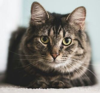 Photo by Kari Shea on Unsplash Photo by Kari Shea on Unsplash Arterial thromboembolism (ATE) in cats has a guarded to poor prognosis and a high mortality/euthanasia rate. In ATE, a blood clot forms in the heart, travels through the circulation, and lodges in an artery. The clot blocks blood flow through that artery, causing significant pain and tissue damage. When the clot blocks an artery supplying a limb, as is usually the case, the limb becomes weak or paralyzed.
The most common site for ATE in cats is the aorta at the point where it divides into smaller arteries that supply the rear legs. A clot stuck here takes on the shape of a saddle, so ATE in this location is called a saddle thrombus. Cats with a saddle thrombus have sudden extreme pain and paralysis of the rear legs (usually both rear legs but sometimes just 1). Witnessing their pain and distress can be quite upsetting to their caretakers. Cause ATE is usually caused by heart disease. Unfortunately, a saddle thrombus can be the very first sign that a cat has heart trouble. The most common type of heart disease in cats—and thus the most common cause of ATE—is hypertrophic cardiomyopathy, an abnormal thickening of the heart muscle. Hypertrophic cardiomyopathy is most common in senior cats but can also affect younger cats. An estimated 15% of all cats and up to 29% of senior cats have hypertrophic cardiomyopathy.[1] Of these cats, a reported 11.3% develop ATE within 10 years after diagnosis.[2] Signs Signs of ATE usually appear suddenly. A cat with ATE will cry loudly in pain and might vomit. The cat will drag the affected limb or be unable to walk. The affected limb might feel cool to the touch. If the cat’s underlying heart disease has progressed to heart failure, the cat might also have signs like panting or rapid breathing. Diagnosis ATE can usually be diagnosed with a physical examination. The affected limb is typically cool, the nail beds are bluer or paler than those of the other paws, and the pulse in that limb is absent or weak. A heart murmur might be present. Diagnostic tests can be used to rule out other possible causes, especially if the cat has less obvious signs caused by a partial blockage. Treatment Many cats with ATE are euthanized before treatment is attempted. Factors linked to a better prognosis include involvement of only 1 limb, ability to move the affected limb, and absence of heart failure (meaning that the heart disease has not yet progressed to the level of heart failure). For cats that receive treatment, the immediate priority is pain control. They also receive anticoagulant medications (blood thinners), treatment for shock and tissue damage, and treatment for heart disease. Cats that don’t have heart failure and survive the first few days in the hospital have a chance of regaining the use of the affected limb once the clot dissolves. Prevention For cats known to have heart disease, certain findings on echocardiography (heart ultrasound) can indicate an increased risk for ATE. These cats can receive preventive anticoagulant medications. However, many cats with heart disease have no obvious signs, so their heart disease isn’t diagnosed. References 1. Luis Fuentes V, Abbott J, Chetboul V, et al. ACVIM consensus statement guidelines for the classification, diagnosis, and management of cardiomyopathies in cats. J Vet Intern Med. 2020;34(3):1062-1077. doi:10.1111/jvim.15745 2. Lo ST, Walker AL, Georges CJ, Li RH, Stern JA. Dual therapy with clopidogrel and rivaroxaban in cats with thromboembolic disease. J Feline Med Surg. 2022;24(4):277-283. doi:10.1177/1098612X211013736 Image source: https://unsplash.com/photos/gray-tabby-cat-FilM6ng7VGQ Laurie Anne Walden, DVM Photo by Anya Prygunova on Unsplash Photo by Anya Prygunova on Unsplash In recent years, monoclonal antibodies have been approved to treat some common medical conditions in animals:
More monoclonal antibodies are likely to become available for dogs and cats. Monoclonal antibodies are used in humans to treat infections, autoimmune disorders, cancer, and other conditions, and they’re being investigated for similar uses in dogs and cats. How Monoclonal Antibodies Work Monoclonal antibodies are in a therapy class called biologics, meaning therapies made from living sources like animal cells, plant cells, or microorganisms. Biologics are large molecules that interact with receptors on the outer surfaces of cells; they don’t enter cells. In contrast, most traditional drugs are small molecules that work inside cells. Monoclonal antibodies are similar to the natural antibodies produced by the immune system. When the immune system detects an antigen, different immune cells release lots of different antibodies that tag different sites on that antigen. (An antigen is anything that triggers an immune response.) Once the antibodies have tagged the antigen, other components of the immune system arrive to destroy it. Monoclonal antibodies are cloned in a laboratory from just one antibody made by one type of immune cell (mono means “one”). They tag only a single site on an antigen. Monoclonal antibody therapy can be very precise because it directs the immune response to a narrow target. In addition to tagging invaders like viruses, monoclonal antibodies can be engineered to tag a receptor on an animal’s own cells. The monoclonal antibodies that relieve pain in dogs and cats, for example, block production of a protein that’s partly responsible for the pain sensation. After monoclonal antibodies have activated the immune system, they’re broken down in the body the same way that natural antibodies are. Unlike small-molecule drugs that enter an animal’s cells, they aren’t cleared through the liver or kidneys. Administration Monoclonal antibodies are given by injection at a veterinary clinic. They can’t be given by mouth because they’re proteins and would be destroyed by digestion. Their effect lasts a few weeks (4-8 weeks for Cytopoint, 4 weeks for Solensia and Librela). These therapies are species specific: monoclonal antibodies for dogs work only for dogs, not for cats. Benefits The monoclonal antibodies that are available for dogs and cats generally work well for their approved uses. They are less likely than some traditional drugs to cause adverse effects because they have a narrow target of action and don’t enter cells. Because they are not cleared through the liver or kidneys, they can be safer than other drugs for animals with liver or kidney disease. Injections given every few weeks might be more convenient for pet owners than pills given multiple times a day, especially if their pets resist oral medication. Adverse Effects Monoclonal antibodies have fewer adverse effects than many traditional drugs. An antibody injection can cause an allergic reaction, although this is rare. Manufacturers might recommend precautions for individual monoclonal antibody products. Image source: https://unsplash.com/photos/a-dog-wearing-a-baseball-glove-E3K7xyAvQ8M Laurie Anne Walden, DVM Photo by Kanashi on Unsplash Photo by Kanashi on Unsplash Vestibular disorders cause problems with balance and coordination. Animals with vestibular disorders typically have a head tilt along with stumbling, walking in circles, and possibly vomiting.
Vestibular problems that start suddenly can look scary to pet owners. The signs can be mistaken for stroke. Some conditions that cause vestibular problems are serious, even life-threatening; others are relatively minor. One of the most common vestibular disorders in dogs is a benign condition called idiopathic vestibular syndrome (idiopathic means “of unknown cause”). This condition is not serious and gets better on its own with time. Idiopathic vestibular syndrome most often affects senior dogs, so in dogs it’s also known as old dog vestibular disease. Cats can also develop idiopathic vestibular syndrome. Causes The vestibular system includes the brainstem, cerebellum (part of the brain), inner ear, and nerves that conduct signals between the inner ear and brain. Anything that affects any part of this system can cause vestibular signs. Vestibular disorders are either central (related to the brainstem and cerebellum) or peripheral (related to the inner ear and surrounding nerves). Central vestibular disorders are usually much more worrisome than peripheral vestibular disorders. Idiopathic vestibular syndrome is a peripheral vestibular disorder. The exact cause in dogs isn’t known. Similar conditions in humans are caused by inflammation of the inner ear or surrounding nerves, viral infections, and benign positional vertigo (an inner ear problem). These same causes are possible but not proven in dogs. Some of the other causes of vestibular disease in animals are deep ear infections (common), cancer, drug adverse effects, toxins, systemic disorders like hypothyroidism, meningitis, and other neurologic disorders. Signs These are the typical signs of a vestibular disorder:
Idiopathic vestibular syndrome usually affects older dogs and cats. It begins suddenly and then starts to improve within a few days. The signs are most pronounced on the first day and don’t get worse with time. In most animals, the signs disappear after a couple of weeks, although the head tilt can last much longer. Other diseases that affect the vestibular system cause signs of their own. For example, a central vestibular disorder might cause seizures or changes in consciousness. Vestibular signs that get worse with time are a signal that the animal has something other than idiopathic vestibular syndrome. Diagnosis Idiopathic vestibular syndrome is a diagnosis of exclusion: it’s diagnosed after other possibilities are ruled out. Diagnosis begins with a neurologic examination, eye and ear examinations, baseline bloodwork, and usually a thyroid function test and urinalysis. Depending on the results, the animal might need referral to a veterinary neurologist and advanced tests like magnetic resonance imaging of the head or cerebrospinal fluid analysis. Treatment For idiopathic vestibular syndrome, the goal of treatment is to make the patient comfortable until the condition resolves on its own. Most animals receive prescription antinausea medication; some also need appetite stimulants or fluid therapy. Home care includes making sure they can eat and drink and protecting them from falling down steps or bumping into things like furniture corners. Physical therapy and postural maneuvers that are used to help people with vertigo aren’t commonly used in animals. Prognosis Most animals with idiopathic vestibular syndrome recover completely, although some have a permanent head tilt afterward. Some animals have more vestibular episodes later on. For vestibular signs that aren’t caused by idiopathic vestibular syndrome, the prognosis depends on whether the cause is treatable. Sources
Image source: https://unsplash.com/photos/brown-and-black-german-shepherd-Xy7SLX9zuVM Laurie Anne Walden, DVM Public domain photo by Ian Livesey on Flickr Public domain photo by Ian Livesey on Flickr Inflammatory bowel disease (IBD) causes chronic inflammation and irritation of the digestive tract. This condition is one of the most common chronic intestinal disorders in cats. IBD in cats is similar to Crohn disease in humans.
Cause In IBD, inflammatory cells accumulate within the walls of the digestive tract. The small intestine is the most common location in cats; the large intestine and stomach can also be affected. The intestinal walls become thickened and can’t absorb nutrients normally. Some cats with IBD also have inflammation of the pancreas and liver, a combination called triaditis. The cause of IBD isn’t known. The disease is probably caused by abnormal immune system function related to intestinal bacteria (which are necessary for normal digestion and immune function), diet, genetics, or other factors. IBD usually affects middle-aged or older cats, although cats of any age can develop the disease. Signs The signs are similar to those of many other digestive system disorders:
Diagnosis Because so many conditions cause the same signs as IBD, cats with chronic digestive tract problems need a thorough workup. Diagnostic tests are performed to rule out a long list of other possibilities, including intestinal lymphoma (a cancer that is clinically very similar to IBD), parasites, intestinal foreign bodies, bacterial and viral infections, food allergies, other types of cancer, and diseases like hyperthyroidism that cause general illness. Baseline tests include fecal parasite tests, bloodwork, and urinalysis. Additional blood tests are used to assess the function of the pancreas and check for evidence that the small intestine is not absorbing nutrients properly. Imaging (radiography and ultrasonography) is used to evaluate the digestive tract, liver, pancreas, and other organs. The only way to definitely diagnose IBD and distinguish it from intestinal lymphoma is to obtain biopsy samples from the intestine and submit them to a diagnostic laboratory for analysis. Biopsy is performed either with endoscopy (tube inserted through the mouth or anus) or abdominal surgery. Both of these procedures require general anesthesia. Treatment IBD is managed, not cured. Treatment is tailored to the individual cat, depending on what works best for that cat. Most cats receive a dewormer to eliminate parasites as a possible cause. Some cats’ signs improve with a hypoallergenic diet, so treatment can include several weeks of a food trial with a prescription diet. Antibiotics are sometimes used carefully (they can disrupt the normal intestinal bacteria). Probiotics and prebiotics, which contain beneficial bacteria or nutrients for existing intestinal bacteria, might be included in the treatment plan. Drugs that suppress the immune response, such as the corticosteroid prednisolone, are necessary for some cats. Prognosis Cats whose disease responds well to treatment can have a good quality of life, although their IBD signs will probably still flare up occasionally. The treatment type and medication doses often need to be adjusted over time. Image source: https://www.flickr.com/photos/ianlivesey/43270035161/ Laurie Anne Walden, DVM Photo by Jonny Neuenhagen on Unsplash Photo by Jonny Neuenhagen on Unsplash The prostate gland grows and develops in response to testosterone and related male sex hormones. Almost all prostate disease that we see in companion animal practice is in intact (not neutered) dogs.
In male animals, neutering means surgical removal of the testicles (castration). Some prostate diseases can affect neutered animals, but these are rare. Most pet male cats in the United States are neutered, so prostate disease is much less common in cats than in dogs. Benign Prostatic Hyperplasia Because the prostate can keep growing as long as it’s exposed to testosterone, most intact male dogs eventually develop enlargement of the prostate, or benign prostatic hyperplasia (BPH). About half of intact male dogs have microscopic evidence of BPH by the time they’re 4 years old; more than 90% have it by the time they’re 8. Most dogs with BPH have no signs of the condition and don’t need treatment. Signs can include blood in the urine, straining to urinate or defecate, and constipation. BPH can reduce the fertility of breeding dogs. In some dogs, an enlarged prostate can be detected with rectal palpation. Ultrasonography is used to measure the prostate and make the diagnosis. Depending on the dog’s signs and test results, samples of prostate fluid or prostate cells might be sent to a laboratory to rule out other prostate disorders. For dogs whose prostate enlargement is causing a problem, the treatment is to eliminate exposure to testosterone, typically with castration. Medications that block the effects of testosterone on the prostate are available for breeding dogs that need to keep their testicles. Prostatitis Dogs with BPH are prone to develop prostatitis, which is infection of the prostate. Prostatitis is fairly common in intact male dogs and rare in neutered male dogs. The signs of prostatitis depend on whether the condition is acute (has just begun) or chronic (has been going on for a long time):
Prostatitis is suspected in any intact male dog with compatible signs—especially if rectal palpation is painful—or repeated urinary tract infections. Diagnostic tests include blood tests, urinalysis, ultrasonography of the prostate, and culture of prostate fluid. Some dogs with prostatitis also have abscesses of the prostate, and these can be seen with ultrasonography. The treatment for prostatitis is several weeks of antibiotics along with treatment of the underlying BPH, which means castration for most dogs. Dogs that are ill with acute prostatitis sometimes need to be hospitalized. Abscesses of the prostate might require surgical drainage. Prostate Cancer Prostate cancer is rare but unfortunately very serious in dogs. The types of prostate cancer that dogs get aren’t linked to testosterone level, so castration does not protect dogs against prostate cancer (it’s actually more common in neutered dogs for unknown reasons). The screening tests and treatments for prostate cancer that are used in men don’t work in dogs. Prostate cancer in dogs tends to be aggressive, is metastatic (spreads through the body), and is usually diagnosed at a late stage. Treatment options other than palliative care are limited; a veterinary oncologist is the best source of advice for an individual dog. Sources Christensen BW. Canine prostate disease. Vet Clin North Am Small Anim Pract. 2018;48(4):701-719. doi:10.1016/j.cvsm.2018.02.012 Palmieri C, Fonseca-Alves CE, Laufer-Amorim R. A review on canine and feline prostate pathology. Front Vet Sci. 2022;9:881232. doi:10.3389/fvets.2022.881232 Image source: https://unsplash.com/photos/black-and-white-short-coated-dog-tKtYHZ13yls Laurie Anne Walden, DVM Public domain photo by Alexandre Burner on Flickr Public domain photo by Alexandre Burner on Flickr Hepatic lipidosis, or fatty liver syndrome, is a life-threatening condition in cats that stop eating for any reason. It’s most common in overweight or obese cats but can affect cats of any weight. Cats that don’t eat for more than a couple of days need to see a veterinarian without further delay.
Causes When cats don’t eat, their bodies start to break down stored body fat to use for energy. Eventually the level of metabolized fat in the blood is higher than the body can use, and the excess fat is stored in the liver cells. The liver cells become swollen with fat and can no longer function normally, resulting in liver failure. The swollen liver cells also squash the bile ducts within the liver, obstructing the flow of bile (which is needed for normal digestion). Overweight and obese cats are at higher risk than average-weight cats because they have more body fat to metabolize. The tendency to store metabolized fat in liver cells is a quirk of feline liver function, so hepatic lipidosis is much more common in cats than in dogs. Anything that makes a cat stop eating can cause hepatic lipidosis. The cause is usually an underlying disease like a digestive system disorder, diabetes, kidney disease, cancer, or hyperthyroidism. Healthy cats can develop hepatic lipidosis if they don’t eat for some reason, like changing to a new food they don’t like, experiencing a stressor like a new pet or a house move, boarding, getting accidentally locked in a garage, getting lost outdoors, and so forth. Signs Cats with hepatic lipidosis usually have signs of liver disease like these:
Cats might also have signs related to the underlying disease that caused them to stop eating. Diagnosis Baseline diagnostic tests (bloodwork and urinalysis) are run to evaluate liver function, assess the cat’s overall condition, and look for an underlying disease. Cats with signs of liver disease usually also have imaging studies like ultrasound of the abdomen. The diagnosis of hepatic lipidosis can be confirmed with liver biopsy, although not all cats need this step. Treatment Cats with hepatic lipidosis are typically hospitalized for at least a few days. Treatment includes intravenous fluids, correction of electrolyte imbalances and anemia (which can happen with liver disease), treatment for nausea and vomiting, nutritional support, and if possible, treatment of the underlying disease. Nutritional support is crucial for cats with hepatic lipidosis so their bodies will stop metabolizing fat for energy. Because most cats with this condition don’t eat on their own, they are fed through a tube at first. Feeding tubes are placed through the nose (cats tolerate this better than you would think) or surgically placed through the skin. Some cats need to continue tube feeding for weeks. These cats receive surgically placed feeding tubes designed for long-term use, and their owners must continue the tube feedings at home. Prognosis Most cats that receive prompt intensive care (including early tube feeding) recover from hepatic lipidosis. The long-term prognosis depends on whether the underlying disease is treatable. Prevention Keeping a cat at a lean body condition reduces the risk of hepatic lipidosis. For cats that stop eating, early intervention to get nutrition into the cat—feeding something the cat will eat, using an appetite stimulant medication, or starting tube feedings—can help prevent hepatic lipidosis. Cats should never be force fed, though. Placing food directly into a cat’s mouth by hand or through a syringe can cause a cat to avoid that food in the future and be even less likely to eat on its own. Image source: https://www.flickr.com/photos/139162177@N05/46079745404/ Laurie Anne Walden, DVM Photo by Alexandre Debiève on Unsplash Photo by Alexandre Debiève on Unsplash Most dogs and cats cope very well with vision loss. They rely more on the sense of smell than on the sense of sight. Blind animals can have happy, comfortable lives with some help from their owners.
Pets with gradual vision loss often adapt so well that their owners don’t realize their pets are having trouble seeing until they’re completely blind and bumping into things. Animals with sudden blindness can take longer to adjust. Safety and Navigation Use gates and other barriers to block a blind animal’s access to stairs, swimming pools, fireplaces, and other dangers. Use foam cushioning (child safety equipment) to pad furniture corners. Keep the floor clear of trip hazards like toys and laundry. Keep blind animals in a crate or other secure space whenever they’re unsupervised, at least while they’re adjusting to their loss of vision. This space can also become a safe, familiar retreat. Blind animals can have a hard time navigating stairs, especially descending. Be patient and use a harness and treats to show blind dogs how to manage stairs. Consider covering short runs of steps with a ramp. Nonslip strips applied to stair treads might make a blind pet feel more secure. Dogs and cats that sleep on furniture might need a ramp or steps so they don’t have to jump up and down. To help blind pets learn their way around the house, don’t pick them up and carry them; let them walk on their own. Different floor surfaces (mats or rugs) can help them identify specific areas like doorways and the location of food and water bowls. A radio left on at all times in one room and scents applied to furniture at the pet’s head height can also help orient them inside the house. Face whiskers help animals feel obstacles, so don’t have their whiskers trimmed at grooming appointments. Vests with circular extensions around the head or chest (halo vests) are available for blind dogs who keep bumping into things. Exercise and Mental Health Blind animals need exercise just like every other animal, but they shouldn’t go outdoors unsupervised. Even a small fenced yard can have holes and fallen branches that would pose a risk to a blind pet. If you don’t have a fenced yard that’s safe and familiar to your dog, keep your dog on a leash. When walking your blind dog, attach the leash to a harness instead of a collar in case you need to pull your dog away from a hazard. Teach your dog verbal cues like “left,” “right,” and “stop.” An unexpected touch can frighten a blind dog, so warn people who approach that your dog is blind. Consider using a dog vest with the words “Blind dog” or “Do not pet.” Be very careful if another dog approaches while you’re walking your dog. A blind dog can’t read other dogs’ body language. If you let your dog interact with another dog, be sure that the other dog is friendly and that your dog is comfortable with the interaction. Provide toys that stimulate senses other than sight. Use toys with bells (with supervision—bells can be a choking hazard), catnip toys for cats, and tracking games for dogs who enjoy following scents. For some animals, playing contact games like tug of war might be easier than chasing toys by sound or scent. Some animals experience anxiety while they’re adjusting to vision loss. Keep your pet’s routine consistent, stay positive around your pet, and provide a safe retreat like a crate. Behavior changes like hiding, reluctance to walk or play, growling, barking, and snapping can all be signs of anxiety. Contact your veterinarian if your pet has any of these changes or seems to be having trouble adapting. Some pets benefit from short-term or long-term prescription anxiety medications. For more ideas, see the ACVO Vision for Animals Foundation website: https://www.visionforanimals.org/coping-with-a-blind-dog/ Image source: https://unsplash.com/photos/SEX4KAz9ExQ |
AuthorLaurie Anne Walden, DVM Categories
All
Archives
June 2024
The contents of this blog are for information only and should not substitute for advice from a veterinarian who has examined the animal. All blog content is copyrighted by Mallard Creek Animal Hospital and may not be copied, reproduced, transmitted, or distributed without permission.
|
- Home
- About
- Our Services
- Our Team
-
Client Education Center
- AKC: Spaying and Neutering your Puppy
- Animal Poison Control
- ASPCA Poisonous Plants
- AVMA: Spaying and Neutering your pet
- Biting Puppies
- Boarding Your Dog
- Caring for the Senior Cat
- Cats and Claws
- FDA warning - Bone treats
- Force Free Alliance of Charlotte Trainers
- Getting your Cat to the Vet - AAFP
- Holiday Hazards
- How To Feed Cats for Good Health
- How to Get the Most Out of your Annual Exam
- Indoor Cat Initiative - OSU
- Introducing Your Dog to Your Baby
- Moving Your Cat to a New Home
- Muzzle Training
- Osteoarthritis Checklist for Cats
- What To Do When You Find a Stray
- Our Online Store
- Dr. Walden's Blog
- Client Center
- Contact
- Cat Enrichment Month 2024
 RSS Feed
|
Office Hours
Monday through Friday 7:30 am to 6:00 pm
|
Mallard Creek Animal Hospital
2110 Ben Craig Dr. Suite 100
|
Site powered by Weebly. Managed by IDEXX Laboratories