Laurie Anne Walden, DVM Intervertebral disk disease (IVDD), also referred to as slipped disk, ruptured disk, or herniated disk, is a relatively common cause of neck and back pain in dogs. Some dogs with IVDD also develop neurologic deficits like toes knuckled under, a wobbly gait, or the inability to walk. The prognosis depends on the severity of the injury and length of time before treatment. A sudden inability to walk is a medical emergency. Affected dogs might need urgent surgery. Anatomy The spine is a row of individual bones called vertebrae. The spinal cord runs along a bony canal through the vertebrae. Intervertebral disks sit between the vertebrae just below the spinal cord and provide cushioning and support for the spine. Intervertebral disks are sort of like jelly doughnuts: they have a squishy interior (the nucleus pulposus) and a firmer, protective exterior (the annulus fibrosus). With IVDD, part of a disk herniates; it leaks or bulges beyond its normal location and presses on the spinal cord or on the surrounding nerves. Types of IVDD IVDD is classified according to the type of disk herniation. Type 1 IVDD has a genetic link: it’s most common in dog breeds with chondrodystrophy, which usually means dogs with short legs and a long back. In type 1 IVDD, the nucleus pulposus becomes mineralized (hardens) over time and eventually breaks through the annulus fibrosus into the spinal canal, bruising the spinal cord. This type of herniation can happen quickly, causing sudden symptoms. Affected dogs are often young adults. Type 2 IVDD is caused by age-related breakdown of the annulus fibrosus. Weakening of the annulus fibrosus allows the disk to bulge upward against the spinal cord. Type 2 IVDD is more gradual than type 1 and is more common in older dogs. Intervertebral disk herniation can also be caused by trauma or strenuous exercise. Dogs at Risk Any dog can develop IVDD. These are the breeds at highest risk for type 1 IVDD:
Type 2 IVDD is more common in larger dogs like these:
Symptoms The symptoms of IVDD range from pain to paralysis, depending on the severity of herniation, the force with which the herniated disk material hits the spinal cord, and the duration of the injury. The mildest form of IVDD is neck or back pain with ability to walk and no neurologic deficits. Neck pain can cause limping, so the symptoms of a herniated disk in the neck can be mistaken for an orthopedic problem. More severe spinal injuries progress through stages that worsen with time: limb weakness, neurologic deficits, inability to walk, inability to stand, inability to move the limbs, and finally inability to feel pain in the limbs. Dogs with spine injuries might lose their normal bladder and bowel function. These are some of the symptoms of IVDD:
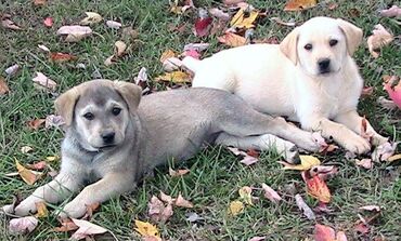
Diagnosis A physical examination shows whether the patient has neurologic deficits and often reveals the approximate location of the injury in the spine. Advanced imaging techniques like computed tomography or magnetic resonance imaging are required to make a definite diagnosis and locate all of the affected disks. Radiographs (x-ray images) are less helpful for diagnosing IVDD but might be obtained to rule out other possible causes of the symptoms, like fractures, dislocations, bone infection, and cancer. Treatment Dogs with milder symptoms—pain, ability to walk, and no or mild neurologic deficits—are usually treated medically at first. A crucial part of medical management is restricting the dog’s activity to allow the annulus fibrosus to heal, similar to treating a sprain. Some dogs need strict crate confinement. Medical management also includes medication to relieve pain. Dogs that begin to feel better with pain medication can reinjure themselves if they resume normal activity too soon, so they usually need to continue strict activity restriction for weeks. Dogs with neurologic deficits or pain that doesn’t improve with medical management often need surgery to remove herniated disk material and relieve pressure on the spinal cord. Time is of the essence; the longer the injury to the spinal cord is present, the worse the neurologic deficits and the worse the prognosis for recovery. For dogs that can’t walk but can still feel sensation in their feet, surgery performed as soon as possible after symptoms begin typically gives the best chance of being able to walk again. Once pain sensation in the feet is lost, the odds of recovery are much lower even with surgery. Spine surgery is expensive and dogs need extensive physical rehabilitation afterward, so the decision to proceed with surgery depends partly on the dog owner’s financial situation and ability to provide long-term care. Sources Olby NJ, Moore SA, Brisson B, et al. ACVIM consensus statement on diagnosis and management of acute canine thoracolumbar intervertebral disc extrusion. J Vet Intern Med. 2022. doi:10.1111/jvim.16480 Spinella G, Bettella P, Riccio B, Okonji S. Overview of the current literature on the most common neurological diseases in dogs with a particular focus on rehabilitation. Vet Sci. 2022;9(8):429. doi:10.3390/vetsci9080429 Photo by PublicDomainPictures on Pixabay Laurie Anne Walden, DVM Photo by C. E. Price Photo by C. E. Price Canine parvovirus is highly contagious among dogs. Infection causes severe illness and is often fatal in dogs that do not receive intensive treatment. Risk Factors Parvovirus infection is most common in puppies that have not yet completed their initial vaccination series and in adult dogs that are not fully vaccinated against parvovirus. A recent outbreak of parvovirus in Michigan highlighted how quickly the virus can spread, especially in dogs in group housing like animal shelters. All dogs are at risk of infection, but whether a dog becomes ill depends on the dog’s level of immunity to the virus. Transmission Canine parvovirus type 2 (the version responsible for illness) infects domestic dogs and some wild animals, such as foxes, coyotes, wolves, and raccoons. The virus is shed in feces and resists drying, heat, and cold, so it stays in the environment for a long time. Dogs are infected by ingesting even a tiny amount of feces that contains parvovirus. Dogs can be infected by contact with an infected dog or with contaminated feces, surfaces, or objects. People who have handled infected dogs or contaminated objects can carry the virus on their hands or clothes and infect other dogs. Wild animals might be a source of environmental spread of parvovirus. Animals that have no symptoms can be carriers. Effects on the Body and Signs of Infection Canine parvovirus targets the lining of the small intestine, bone marrow, heart, and other organs. Destruction of the intestinal lining causes vomiting, bloody diarrhea, dehydration, loss of barrier protection against bacteria, and inability to absorb nutrients. Some infected animals develop intussusception, a potentially life-threatening condition in which a section of intestine folds into itself like a telescope. White blood cells (part of the immune system) are produced in the bone marrow, so infected animals have low white blood cell counts and impaired immune function. Because of decreased intestinal barrier protection and immune function, infected animals are at risk of septic shock caused by bacterial infection. Young puppies sometimes develop myocarditis, or inflammation of the heart muscle. These are some of the signs of parvovirus infection:
Death can occur within 3 days after symptoms begin. Diagnosis Parvovirus infection is often suspected on the basis of the patient’s symptoms, age, and vaccination history. Tests of fecal samples confirm the infection. A rapid test at a veterinary clinic is convenient but sometimes returns a false result, which can delay the diagnosis (this was the case with the outbreak in Michigan). A polymerase chain reaction (PCR) test at a diagnostic laboratory is accurate but takes longer. A blood test showing a low white blood cell count supports a diagnosis of parvovirus, especially in a sick puppy still awaiting a PCR test result. Dogs with parvovirus infection usually also receive other tests, such as x-ray imaging or ultrasound of the belly. Treatment For most dogs with parvovirus infection, the best chance for recovery is hospitalization for intensive care very soon after symptoms start. No antiviral drug that kills parvovirus is currently available, so treatment consists of measures to reverse dehydration, minimize vomiting and diarrhea, prevent bacterial infection, reduce pain, and restore nutrition. Intensive care for parvovirus infection can be expensive. Prognosis Most dogs that receive prompt intensive treatment in the hospital survive. Most dogs that are not treated die. For dogs whose owners can’t afford hospitalization, the success of outpatient treatment depends on the individual dog (illness severity and immune status), type of treatment, and ability of the owners to provide care at home. Prevention The parvovirus vaccine is effective and is one of the core vaccines recommended for all dogs. This vaccine is part of the initial series of puppy vaccines. Puppies need to receive the entire initial vaccine series to have the best chance of being protected. The current recommendation is for puppies to be vaccinated every 2 to 4 weeks from the ages of about 6 weeks to at least 16 weeks. The vaccine is given again 1 year later and then every 3 years throughout the dog’s life. Some dogs never mount an immune response to vaccination (this is not common). Vaccinating all dogs helps keep puppies and vulnerable adult dogs safe. Limiting a puppy’s exposure to other dogs and to places where other dogs gather reduces the risk of infection. However, this approach can hinder a puppy’s social and emotional development. In general, puppy classes and establishments that have protocols to reduce disease transmission (like requiring vaccines, proper disinfection and hygiene, and isolation of sick dogs) are relatively safe, especially when weighed against the risk of behavior problems in puppies that are not socialized. Exposure to dogs of unknown vaccination and health status, as in dog parks, is riskier. Ask your veterinarian about safe activities for your puppy, and keep your puppy away from sick dogs and other dogs’ feces. For More Information
Public domain photo by C. E. Price |
AuthorLaurie Anne Walden, DVM Categories
All
Archives
June 2024
The contents of this blog are for information only and should not substitute for advice from a veterinarian who has examined the animal. All blog content is copyrighted by Mallard Creek Animal Hospital and may not be copied, reproduced, transmitted, or distributed without permission.
|
- Home
- About
- Our Services
- Our Team
-
Client Education Center
- AKC: Spaying and Neutering your Puppy
- Animal Poison Control
- ASPCA Poisonous Plants
- AVMA: Spaying and Neutering your pet
- Biting Puppies
- Boarding Your Dog
- Caring for the Senior Cat
- Cats and Claws
- FDA warning - Bone treats
- Force Free Alliance of Charlotte Trainers
- Getting your Cat to the Vet - AAFP
- Holiday Hazards
- How To Feed Cats for Good Health
- How to Get the Most Out of your Annual Exam
- Indoor Cat Initiative - OSU
- Introducing Your Dog to Your Baby
- Moving Your Cat to a New Home
- Muzzle Training
- Osteoarthritis Checklist for Cats
- What To Do When You Find a Stray
- Our Online Store
- Dr. Walden's Blog
- Client Center
- Contact
- Cat Enrichment Month 2024
|
Office Hours
Monday through Friday 7:30 am to 6:00 pm
|
Mallard Creek Animal Hospital
2110 Ben Craig Dr. Suite 100
|
Site powered by Weebly. Managed by IDEXX Laboratories

 RSS Feed