Laurie Anne Walden, DVM Photo by Sophie Shankey on Unsplash Photo by Sophie Shankey on Unsplash Tracheal collapse causes a chronic dry cough that sounds similar to a goose honk. The disease tends to get worse with time. Tracheal collapse can’t be cured, but for most patients, medical treatment reduces the severity of the cough. In the most serious cases, tracheal collapse interferes with breathing and requires emergency care.
Tracheal collapse can affect dogs of any breed but is most common in small-breed dogs like Yorkshire terriers and Pomeranians. It’s rare in cats. Cause The trachea (windpipe) is a tube held open by rings of cartilage. In animals with tracheal collapse, the cartilage rings soften and become weakened over time. The cartilage rings lose their ability to hold the trachea open and the trachea flattens during breathing or coughing, blocking the flow of air. The severity of the signs depends on the percentage of airway that’s blocked. The signs also partly depend on the location of the affected section of trachea: outside the rib cage (in the neck) versus within the rib cage. In dogs with tracheal collapse, coughing is made worse by pressure on the neck, excess weight, stress, excitement, exercise, respiratory irritants like smoke, and respiratory infections. Some dogs with tracheal collapse also have abnormalities of the larynx and the bronchi (airways inside the lungs). Chronic coughing causes airway inflammation, which in turn leads to more coughing. Tracheal collapse and heart disease are both common in middle-aged and older small-breed dogs, so many dogs have both diseases at the same time. Both diseases cause coughing and exercise intolerance. Figuring out which disease is most responsible for the cough can be a bit of a diagnostic challenge. Signs
Diagnosis Tracheal collapse is suspected on the basis of the patient’s history, the sound of the cough, and physical examination findings. A definite diagnosis is made with imaging studies. Radiographs (x-ray images) sometimes show a collapsed trachea and are also used to evaluate heart size and the appearance of the lungs. In many dogs the trachea collapses only on inhalation or only on exhalation, so fluoroscopy—an x-ray “movie”—can reveal collapse that doesn’t appear on radiographs. Tracheoscopy is examination of the inside of the trachea and bronchi with a fiber optic camera in an endoscopy tube. An advantage of tracheoscopy is that it allows samples from inside the trachea to be collected for laboratory analysis; a disadvantage is that it requires general anesthesia. Dogs with tracheal collapse might have bloodwork to assess their overall condition, especially if they are older or have concerning clinical signs. Because the signs of tracheal collapse and heart disease overlap, dogs with suspected or known heart disease also benefit from echocardiography to evaluate heart function. Treatment Tracheal collapse isn’t curable, so medical treatment needs to continue for life. Medications chosen for an individual patient might include cough suppressants to break the cough cycle, corticosteroids to reduce airway inflammation, bronchodilators, and antianxiety medications or sedatives to reduce stress. The choice of medication can change over time as the dog’s disease progresses. Nonmedical management is crucial for dogs with tracheal collapse and includes the following:
Surgical procedures like tracheal stent placement are available for dogs with severe signs. However, surgical options don’t cure the underlying tracheal problem and might not eliminate the cough. Image source: https://unsplash.com/photos/a-small-dog-standing-under-a-wooden-bench-HSs4t4TWPFg Laurie Anne Walden, DVM Toxoplasma gondii oocysts in fecal flotation solution. Image from CDC. Toxoplasma gondii oocysts in fecal flotation solution. Image from CDC. Have you ever heard that pregnant people shouldn’t handle a cat’s litter box? This is because of the risk of toxoplasmosis. World Zoonoses Day, July 6, is a great time to talk about how this disease passes from animals to people and whether pregnant people really do get a 9-month pass on cleaning the litter box.
Zoonoses are diseases that are transmitted from nonhuman animals to humans. Toxoplasmosis is a zoonotic disease caused by infection with the protozoan parasite Toxoplasma gondii. Toxoplasmosis is one of the most common foodborne illnesses in people. The people who should be most cautious about T gondii infection are those with compromised immune systems and those who are pregnant. Toxoplasmosis in People People With Healthy Immune Systems People with healthy immune systems usually have no signs of T gondii infection and don’t know they’ve been infected. For those who develop signs, most have swollen lymph nodes or flu-like symptoms that get better on their own within a few weeks. Much less commonly, the infection can affect the eyes and cause vision loss. Whether or not the initial infection causes any signs of illness, the parasites remain in the body in a dormant state. The infected person develops immunity to T gondii. However, if the person’s immune system becomes compromised (because of chemotherapy, organ transplantation, or AIDS, for example), the T gondii organisms are reactivated and cause illness. People With Weakened Immune Systems In people with compromised immunity, toxoplasmosis affects the brain, eyes, lungs, and other organs. Either a new infection or a reactivated infection can cause seizures, confusion, and coordination problems. Fetuses Infected Before Birth A fetus won’t become infected if the mother was infected with T gondii before getting pregnant. In this case the mother already has immunity and won’t pass the infection to the unborn child (as long as she has a healthy immune system). If a mother is infected for the first time during or immediately before pregnancy, the fetus can be infected too because the mother doesn’t yet have immunity. Most people don’t know if they’ve been infected with T gondii in the past, so it’s safest for all pregnant people to take precautions to avoid infection. The consequences of infection in an unborn child can be severe. The pregnancy might end in miscarriage or stillbirth, or the fetus might develop anatomic abnormalities. More commonly, infected infants have no signs of infection when they’re born. These individuals can develop vision loss or neurologic problems years later. How People Are Infected
Toxoplasmosis in Cats Domestic cats and other felines are the only species that spread T gondii into the environment. Cats become infected after eating birds or rodents that have T gondii cysts in their bodies. Infected cats then shed T gondii oocysts (eggs) in the stool. The oocysts are not infective to other animals right away. After at least 1 day, the oocysts mature to a stage that can infect other animals. Infective oocysts can survive in the environment for years. Most infected cats don’t have any signs of illness. Cats with compromised immune systems can develop disease similar to that of humans with compromised immune systems. Identifying cats infected with T gondii is tricky. Cats shed oocysts in the stool for only a short time, so fecal tests aren’t reliable for diagnosing infection in cats. However, if a fecal test happens to show oocysts that look similar to T gondii, the cat’s owners should take precautions with the cat’s stool for a few weeks. A blood test showing that a cat has T gondii antibodies doesn’t tell us whether that cat is likely to start shedding oocysts that could put the family at risk. Precautions The CDC recommends these precautions to avoid toxoplasmosis:
Pregnant people should also take these precautions:
For More Information
Image source: https://www.cdc.gov/dpdx/toxoplasmosis/index.html Laurie Anne Walden, DVM Photo by Madalyn Cox on Unsplash Photo by Madalyn Cox on Unsplash Environmental enrichment means modifying an animal’s living space to encourage physical activity and allow the animal to engage in behaviors that are natural for its species. For indoor pets especially, environmental enrichment improves physical and mental health and can reduce behavior problems.
Dogs and cats with mobility problems need environmental modification for accessibility and mental stimulation. Pets have mobility limitations for a variety of reasons:
Household Modifications and Physical Activity Cover hard floors with nonslip mats or rugs. Consider using lots of small washable mats instead of large rugs for easier cleaning if your pet urinates or defecates on the floor (common in animals with medical problems, mobility limitations, or cognitive decline). Be sure to have a nonslip surface in front of your pet’s food and water bowls. Use steps and ramps. Think of all the places your pet likes to be that are not the floor—bed, sofa, cat tree (climbing structure), window seat, and so forth—and make sure your pet can get up and down without jumping. You might need to train your pet to use steps and ramps by leading them with treats. Cats need to scratch for claw health and their mental health (it’s normal species behavior). Reaching and stretching can be difficult for cats with arthritis, so offer your cat horizontal as well as vertical scratching surfaces. Ask your veterinarian about exercise that’s appropriate for your pet. Depending on the medical condition, your pet might benefit from controlled gentle exercise. Some veterinary facilities have physical rehabilitation equipment like underwater treadmills. Toileting Difficulty reaching the toileting area causes anxiety for animals. For cats, put low-sided litter boxes on every floor of the house in quiet, accessible areas. Cats tend to be most comfortable toileting in big open boxes (at least 1.5 times the length of the cat, not including the tail). A large plastic storage bin makes a great litter box; cut an opening on one side so your cat can walk in and out easily. Dogs with new mobility problems or cognitive decline might not give you their usual cues that they need to go out. If your dog or cat starts having toileting accidents indoors, make a veterinary appointment to check for a medical reason. If no medical cause is found, your pet might be having trouble getting outside or to the litter box. Mental Stimulation Engage your pet’s senses: use toys with different odors, textures, and sounds. Puzzle feeders and treat-dispensing toys engage the brain and encourage physical activity. Teach your old dog or cat new tricks. Use positive reinforcement to train new behaviors that don’t require a lot of movement (like touching their nose to your hand). For dogs with mobility problems, walks aren’t really about walking. Just seeing and smelling the outdoors is valuable mental enrichment for dogs. Take your dog out on a harness and forget about actually getting anywhere; be patient and let him sniff and wander at his own pace. If your dog can’t walk easily, carry him or use a wagon. Give your cat a soft seat by a window if possible. Your cat might also enjoy cat-friendly videos. For More Ideas Home modifications. International Cat Care. 2024. Accessed June 18, 2024. https://icatcare.org/app/uploads/2024/03/Changes-to-the-home-environment_ISFM-caregiver-guide_FINAL.pdf Sueda K, Cho J. Environmental enrichment for senior dogs and cats. Clinician’s Brief. December 2017. Accessed June 18, 2024. https://www.cliniciansbrief.com/article/environmental-enrichment-senior-dogs-cats Image source: https://unsplash.com/photos/black-cat-on-white-cat-tree-X7OokuRyvCI Laurie Anne Walden, DVM Photo by Saru Robert on Unsplash Photo by Saru Robert on Unsplash If your cat vomits, don’t assume it’s because of hairballs (or just because he’s sitting on an expensive rug). Vomiting has lots of possible causes, and some are serious. A mistaken belief that vomiting is normal for cats can delay diagnosis and treatment.
When to Seek Veterinary Care Many cats vomit occasionally, just like dogs and people. Some cats vomit frequently, and these cats need an examination and diagnostic tests. “Frequently” means different things for different cats, but in general, cats that vomit more than about once a month should see a veterinarian. These are some other signs that a vomiting cat needs veterinary care:
Vomiting or Coughing? Respiratory problems in cats can be mistaken for vomiting. Coughing can look a lot like hacking up a hairball. Asthma is common in cats, and the hunched posture and raspy breathing of an asthma attack can look and sound very similar to retching. True vomiting means forcefully expelling stomach contents from the mouth. Hacking up food or a hairball is vomiting. Hacking up a little bit of foam is harder to interpret. Signs that the problem is with the lungs or heart instead of the stomach include open-mouth breathing, wheezing, and increased sleeping respiratory rate (more than 30 breaths per minute while asleep). It’s OK if you can’t tell if your cat is vomiting or coughing. Take a video to show your veterinarian and make an appointment as soon as you can; lung and heart problems shouldn’t wait. Causes of Vomiting Vomiting has too many possible causes to list here. These are some of the most common in cats:
Diagnostic Tests The tests chosen depend on the cat’s history, signs, and physical examination findings. Cats with chronic vomiting typically undergo a series of tests until the cause is found. Baseline laboratory tests include bloodwork, urinalysis, and stool tests for parasites. Radiography (x-ray imaging) can show some of the problems that cause vomiting; ultrasonography is more useful for others. Inflammatory bowel disease and some other intestinal disorders are diagnosed with biopsy of the intestine, which requires general anesthesia. Treatment The type and duration of treatment depend on the cause. Foreign objects and hairballs that are blocking the intestine are removed surgically. Some problems can be treated with a single course of medication; others require lifelong treatment. If your cat resists oral medication (pills or liquids), tell your veterinarian so you can explore alternatives. Image source: https://unsplash.com/photos/a-gray-cat-sitting-on-top-of-a-rug-ybibgcEcv2w Rachel Gutierrez, DVM Cats need access to food, water, shelter, and litter boxes. We can consider these their essential needs to survive; however, they also need means to express typical species behavior like hunting, viewing, climbing, scratching, and playing. Enrichment enhances our cat’s environment by giving them chances to express these behaviors. This can improve their mental and physical well-being and even reduce unwanted behaviors. Any changes in routine can be stressful for cats and any new activity or changes in environment are recommended to be done slow and steady.
Different Types of Enrichment Food puzzles/foraging toys: Food puzzles are a great way to increase your cat's activity and mental stimulation. There are a variety of commercially made food puzzles like Doc and Phoebe’s mice, sniffle mats, Trixie puzzle feeders, etc. Some of these are stationary and others cats have to move around to get food out of them. You can also make your own using cardboard boxes, towels, toilet paper roll, etc. When introducing your cat to any food puzzle, recommend starting easy then building up to more challenging puzzles. Toys: Cats have a natural instinct to hunt and we can use this instinct to get them to play. Cats typically like mice, bugs, and birds as prey when they live outdoors. With this in mind, using toys that mimic the feel, sound, or movement of mice, bugs, or birds can entice cats to play more. Some examples are wand toys with feathers or faux fur OR cat dancer toys (mimic a bug). It is best to rotate toys so they do not lose interest (ideally every few days). Clicker training: Cats can be trained to do a variety of tricks (sit, high-five, lay down, etc) or go through an obstacle course. Clicker training can even be used to help you medicate your cat (with appropriate training). Recommend getting a clicker with a ball at the end (this can be purchased online). Sensory enrichment: Videos of mice, birds, or fish can be stimulating for cats (YouTube videos). Or set up a bird feeder outside near their favorite window with a window perch. For their sense of smell and need to chew, catnip or silvervine or cat grass can be stimulating for them. Outdoor walks or screened-in porches are great sources of enrichment for cats as well (please ensure this is under supervision). Environmental enrichment (cat trees, scratchers, and more): One of the cat’s essential needs is shelter, which includes a safe place to sleep and rest; however, this also includes a place to have time alone or get away from people or other animals. This is where cat trees or other vertical spaces can be very useful. It also creates areas where you can have playtime or feed them or use it as part of an obstacle course. Another important behavior/need cats have is scratching. This is important for their nail health. Many cats have preferences of the material and vertical versus horizontal. I recommend trying out the different options. There are a variety of other cat furniture ideas you can use for enrichment, such as shelves, tunnels, window perches, etc. Consider changing the positions or locations of any of these to renew their interest. Check out these resources for more ideas regarding feline enrichment:
Laurie Anne Walden, DVM Photo by Manja Vitolic on Unsplash Photo by Manja Vitolic on Unsplash Urinary tract disorders are common in cats. Feline lower urinary tract disease (FLUTD) is a general term that describes disorders of the bladder and urethra. A number of disorders cause FLUTD, so cats with any changes in urination or litter box behavior need to see a veterinarian.
Signs Cats with FLUTD have similar signs regardless of the underlying cause:
Causes Idiopathic Cystitis The most common cause of FLUTD is idiopathic cystitis. Idiopathic means “of unknown cause,” so this condition is diagnosed when tests don’t reveal another cause. Chronic stress appears to be a major risk factor. An abnormality of the bladder lining could also be involved. Idiopathic cystitis is managed rather than cured. The signs tend to come and go over time. Flare-ups often improve on their own regardless of treatment, which makes it hard to tell if a particular treatment has actually had any effect. Cats with idiopathic cystitis are treated with stress-reducing measures like environmental enrichment and antianxiety medication. Some cats benefit from other medications or special diets. Urinary Tract Infection Urinary tract infections are uncommon in young and middle-aged cats. Cat urine is normally so concentrated that bacteria can’t easily survive in it. However, diseases that affect urine concentration and chemical composition (like kidney disease and diabetes) are very common in older cats, so cats in this age group are much more likely to have urinary tract infections. A diagnosis of urinary tract infection requires urinalysis and ideally a urine culture to identify the specific bacteria. Older cats with urinary tract infections should also have bloodwork and radiography or ultrasonography of the abdomen to find the underlying cause. Bacterial urinary tract infections are treated with antibiotics, ideally chosen according to culture results. Uroliths (Bladder Stones) Uroliths are rocklike mineral collections that form in the bladder or any other part of the urinary tract. Uroliths are found with radiography or ultrasonography. Some types of uroliths can be dissolved with a prescription diet; others require surgical removal. Even the dissolvable types often need to be removed surgically because they can be painful, cause infection, and block the urethra of male cats. Uroliths that form in a kidney or ureter (the tube connecting the kidney to the bladder) usually warrant referral to a veterinary surgery specialist. Urethral Obstruction Urethral obstruction is a life-threatening condition in which a urolith or mucus plug blocks the urethra. Male cats are at much higher risk than female cats because male cats’ urethras are narrower and longer. Cats with urethral obstruction initially have the same signs as cats with FLUTD from any other cause. If the obstruction isn’t removed, the bladder becomes large and painful and can rupture. The kidneys can no longer function normally, and the cat can die of toxin buildup and electrolyte imbalance. Cats with urethral obstruction need emergency care and usually at least a few days of hospitalization. Other Causes Cancer, injury of the spinal cord or urinary tract, and anatomical problems with the urinary tract can also cause FLUTD. Image source: https://unsplash.com/photos/black-and-white-cat-lying-on-brown-bamboo-chair-inside-room-gKXKBY-C-Dk Laurie Anne Walden, DVM Baby striped skunk. Photo by Kevin VanGorden, USDA Forest Service. Baby striped skunk. Photo by Kevin VanGorden, USDA Forest Service. Skunks aren’t aggressive, but they’ll spray dogs that get too close. Skunk spray is a smelly nuisance that doesn’t usually cause medical problems. However, it’s irritating to the eyes and in rare cases can cause more serious problems.
Dogs are most likely to encounter skunks during warm months. In North Carolina, skunk breeding season is February through April; kits are born in May and June. Skunks live in cities like Charlotte, not just in rural areas. You might not see them often—or at all—because they’re most active at night. They live in wooded or brushy areas and raid garbage cans in residential neighborhoods. Skunk Spray Skunk spray is thick, oily material secreted from the anal glands. The nasty smell comes from thioacetates and thiols, chemical compounds that contain sulfur—the same element that makes rotten eggs so stinky. Thioacetates (the less smelly compounds) are converted to thiols (super smelly) when they’re exposed to water, and sulfur atoms tend to bind tightly to other atoms, so removing skunk odor from fur can be difficult. Because water activates the thiol conversion, a dog that’s been skunked can smell skunky after baths for a long time. Skunks can control their anal glands in ways that dogs and cats can’t. They can aim their anal gland spray at a target several feet away, or they can release a cloud of mist. Baby skunks can spray targets too. Risks to Dogs Spray Skunk spray in the eyes can cause redness, eyelid swelling, corneal damage, and temporary blindness. Inhaled skunk spray irritates the respiratory tract. The spray can also cause nausea and vomiting. Dogs sprayed by a skunk can develop life-threatening hemolytic anemia, although fortunately this is rare. The compounds in skunk spray can destroy red blood cells. The risk is likely highest for dogs that swallow the spray, dogs exposed to a large volume, and dogs with multiple exposures. Dogs with hemolytic anemia often need to be hospitalized at a 24-hour clinic and might need a blood transfusion. Bite Wounds and Rabies Skunks are a relatively common rabies carrier species in North Carolina. However, the chance that an individual skunk has rabies is low. Rabies is spread through saliva, not through skunk spray. Most dogs that encounter a skunk are sprayed before they get close enough to be bitten. Still, any dog that has been near a skunk should be checked carefully for bite wounds. Dogs with a bite wound need to see a veterinarian immediately for wound care and assessment of rabies risk. In North Carolina, dogs with a possible rabies exposure (for example, a bite wound from a rabies carrier species) need a rabies vaccine booster within 96 hours. What to Do if Your Dog Gets Skunked Most dogs sprayed by skunks don’t need to see a veterinarian. Monitor your dog for a few days and seek veterinary care if your dog has any of the following:
Deskunk your dog:
Prevention Keep your dog’s rabies vaccine up to date and make your yard unattractive to skunks:
For More Information
Image source: https://www.flickr.com/photos/lblkytn/14928066475/ Laurie Anne Walden, DVM Photo by Kari Shea on Unsplash Photo by Kari Shea on Unsplash Arterial thromboembolism (ATE) in cats has a guarded to poor prognosis and a high mortality/euthanasia rate. In ATE, a blood clot forms in the heart, travels through the circulation, and lodges in an artery. The clot blocks blood flow through that artery, causing significant pain and tissue damage. When the clot blocks an artery supplying a limb, as is usually the case, the limb becomes weak or paralyzed.
The most common site for ATE in cats is the aorta at the point where it divides into smaller arteries that supply the rear legs. A clot stuck here takes on the shape of a saddle, so ATE in this location is called a saddle thrombus. Cats with a saddle thrombus have sudden extreme pain and paralysis of the rear legs (usually both rear legs but sometimes just 1). Witnessing their pain and distress can be quite upsetting to their caretakers. Cause ATE is usually caused by heart disease. Unfortunately, a saddle thrombus can be the very first sign that a cat has heart trouble. The most common type of heart disease in cats—and thus the most common cause of ATE—is hypertrophic cardiomyopathy, an abnormal thickening of the heart muscle. Hypertrophic cardiomyopathy is most common in senior cats but can also affect younger cats. An estimated 15% of all cats and up to 29% of senior cats have hypertrophic cardiomyopathy.[1] Of these cats, a reported 11.3% develop ATE within 10 years after diagnosis.[2] Signs Signs of ATE usually appear suddenly. A cat with ATE will cry loudly in pain and might vomit. The cat will drag the affected limb or be unable to walk. The affected limb might feel cool to the touch. If the cat’s underlying heart disease has progressed to heart failure, the cat might also have signs like panting or rapid breathing. Diagnosis ATE can usually be diagnosed with a physical examination. The affected limb is typically cool, the nail beds are bluer or paler than those of the other paws, and the pulse in that limb is absent or weak. A heart murmur might be present. Diagnostic tests can be used to rule out other possible causes, especially if the cat has less obvious signs caused by a partial blockage. Treatment Many cats with ATE are euthanized before treatment is attempted. Factors linked to a better prognosis include involvement of only 1 limb, ability to move the affected limb, and absence of heart failure (meaning that the heart disease has not yet progressed to the level of heart failure). For cats that receive treatment, the immediate priority is pain control. They also receive anticoagulant medications (blood thinners), treatment for shock and tissue damage, and treatment for heart disease. Cats that don’t have heart failure and survive the first few days in the hospital have a chance of regaining the use of the affected limb once the clot dissolves. Prevention For cats known to have heart disease, certain findings on echocardiography (heart ultrasound) can indicate an increased risk for ATE. These cats can receive preventive anticoagulant medications. However, many cats with heart disease have no obvious signs, so their heart disease isn’t diagnosed. References 1. Luis Fuentes V, Abbott J, Chetboul V, et al. ACVIM consensus statement guidelines for the classification, diagnosis, and management of cardiomyopathies in cats. J Vet Intern Med. 2020;34(3):1062-1077. doi:10.1111/jvim.15745 2. Lo ST, Walker AL, Georges CJ, Li RH, Stern JA. Dual therapy with clopidogrel and rivaroxaban in cats with thromboembolic disease. J Feline Med Surg. 2022;24(4):277-283. doi:10.1177/1098612X211013736 Image source: https://unsplash.com/photos/gray-tabby-cat-FilM6ng7VGQ Laurie Anne Walden, DVM Photo by Rafaëlla Waasdorp on Unsplash Photo by Rafaëlla Waasdorp on Unsplash All dogs are at risk for leptospirosis and should be vaccinated for it every year, according to updated recommendations from the American College of Veterinary Internal Medicine (ACVIM).
Leptospirosis is caused by infection with Leptospira bacteria. The infection spreads from animals to humans, so leptospirosis is a public health hazard. Leptospirosis is a serious disease that damages the kidneys and other organs. The leptospirosis vaccine for dogs has previously been considered a lifestyle vaccine, given to dogs with certain risk factors but not necessarily to all dogs. Information about how the disease spreads has improved over the years, so the ACVIM now says that all dogs, regardless of their lifestyle and location, should receive the vaccine. Transmission Leptospira spread mainly through urine. Many wild and domestic animal species carry the bacteria and deposit them in the environment. Rats and other rodents are the most common carriers. Leptospira grow best in water and wet soil. Dogs with access to bodies of water, especially in warm climates, are at higher risk than others. However, the infection can also spread by direct contact (for example, when dogs hunt rodents) and in other ways, so outbreaks have happened among people and dogs living in urban areas and in drier and colder climates. Cats seem to be resistant to leptospirosis, and cases in cats are extremely rare. However, cats might be able to spread the bacteria to other animals. Disease Leptospirosis affects many organ systems. These are some of the most serious problems it causes:
These are some of the signs of infection:
Diagnosis and Treatment Leptospira can be hard to detect with laboratory tests. Definite diagnosis might require multiple blood and urine tests to isolate bacterial DNA and measure antibody titers. Dogs with suspected leptospirosis also receive baseline bloodwork, urinalysis, and chest radiographs. Leptospirosis is treated with antibiotics. Additional treatments depend on the individual patient’s course of illness and the organ systems affected. Some dogs with kidney injury caused by leptospirosis need dialysis. The risk of a person contracting leptospirosis directly from an infected dog is low because dogs don’t shed many Leptospira organisms in their urine. However, people caring for a dog with leptospirosis need to take precautions like avoiding contact with the urine, wearing protective clothing, and washing hands. Vaccination The doctors at Mallard Creek recommend that all dogs receive a leptospirosis vaccine starting at 9 weeks of age. Dogs need a booster vaccine 3 to 4 weeks later and then once a year. Leptospirosis vaccines are not all the same. Leptospira have many different variations, or serovars, and vaccines target only specific serovars. However, some don’t cover the serovars currently causing disease in dogs in the United States. Your veterinarian can advise you on the best vaccination plan for your own dog. Source Sykes JE, Francey T, Schuller S, Stoddard RA, Cowgill LD, Moore GE. Updated ACVIM consensus statement on leptospirosis in dogs. J Vet Intern Med. 2023;37(6):1966-1982. doi:10.1111/jvim.16903 Image source: https://unsplash.com/photos/a-dog-running-through-a-field-of-yellow-flowers-SEGsw2Kmd08 Laurie Anne Walden, DVM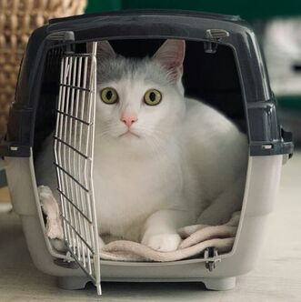 Photo by Anatolii Kozhukhar on Unsplash Photo by Anatolii Kozhukhar on Unsplash Many dogs and cats experience fear, anxiety, and stress at the veterinary clinic. Their anxiety often begins before they even arrive at the clinic, especially if they associate pet carriers and car rides with the clinic. Previsit antianxiety medications for pets can make clinic visits easier and safer for these pets, their owners, and clinic staff.
These are some of the benefits of previsit medications for anxious pets:
Previsit medications have benefits for pet owners and clinic staff members too. Being the caretaker of an anxious and fearful pet isn’t easy, so previsit medications for pets can reduce everyone’s stress levels. For clinic staff members, managing patients’ clinic anxiety reduces the risk of injury and decreases workplace stress (we love animals too and don’t want to cause them distress). How to Know if Your Pet Needs Previsit Medications Previsit medications aren’t just for aggressive animals. Pets with more subtle signs of stress are scared too, and they deserve help for their anxiety just as much as animals that can’t be handled without full sedation. If your pet shows obvious signs of fear at the clinic, you probably already know that your pet would benefit from previsit medications. But maybe your pet’s signs are not as noticeable and you’re a little surprised when your veterinarian recommends medications. Dogs and cats show anxiety and fear with a range of behaviors and body language cues. Anxious and fearful dogs and cats tend to start with subtle behaviors like lip licking or freezing. If the scary thing doesn’t go away—or tries to draw a blood sample—the behavior can escalate to aggression. Dogs and cats that are anxious at the veterinary clinic can show these signs:
How to Use Previsit Medications Previsit medications work best if they’re used along with training and positive reinforcement. For example, a cat can make positive associations with the cat carrier if the carrier is left out all the time as part of the normal household furniture, sometimes with yummy treats inside, instead of appearing only when the cat is going to the clinic. Veterinarians choose previsit medications according to the needs and medical condition of the individual patient. Some of the drugs commonly prescribed for situational (short-term) anxiety are gabapentin, trazodone, and clonidine. Motion sickness contributes to anxiety, so pets might also receive antinausea medication. Some of these medications can cause sedation, but the main goal is anxiety reduction, not sedation. Previsit medications are timed to have maximum effect at the time of the clinic visit. They need to take effect before the pet experiences any stress. Most of these medications are given at least 1 to 2 hours before the clinic visit; sometimes a loading dose is also given the night before. They typically last for up to 8 hours, although the duration can vary. Because individual animals respond differently to these medications, giving a trial dose can be very helpful. If you use a trial dose, tell your veterinarian how long it took for the medication to take effect, how the medication affected your pet’s behavior, whether your pet had adverse effects like vomiting or excessive sedation, and how long the medication’s effects lasted. For some dogs and cats, previsit medications aren’t enough to overcome their fear. These pets might also need a sedative injection at the clinic. For these pets, previsit medications given at home make the sedative injection easier for the pet (and staff) and can also reduce the dose of injectable drug. Image source: https://unsplash.com/photos/a-white-cat-sitting-inside-of-a-cage-B-_SyRxv2So |
AuthorLaurie Anne Walden, DVM Categories
All
Archives
June 2024
The contents of this blog are for information only and should not substitute for advice from a veterinarian who has examined the animal. All blog content is copyrighted by Mallard Creek Animal Hospital and may not be copied, reproduced, transmitted, or distributed without permission.
|
- Home
- About
- Our Services
- Our Team
-
Client Education Center
- AKC: Spaying and Neutering your Puppy
- Animal Poison Control
- ASPCA Poisonous Plants
- AVMA: Spaying and Neutering your pet
- Biting Puppies
- Boarding Your Dog
- Caring for the Senior Cat
- Cats and Claws
- FDA warning - Bone treats
- Force Free Alliance of Charlotte Trainers
- Getting your Cat to the Vet - AAFP
- Holiday Hazards
- How To Feed Cats for Good Health
- How to Get the Most Out of your Annual Exam
- Indoor Cat Initiative - OSU
- Introducing Your Dog to Your Baby
- Moving Your Cat to a New Home
- Muzzle Training
- Osteoarthritis Checklist for Cats
- What To Do When You Find a Stray
- Our Online Store
- Dr. Walden's Blog
- Client Center
- Contact
- Cat Enrichment Month 2024
 RSS Feed
|
Office Hours
Monday through Friday 7:30 am to 6:00 pm
|
Mallard Creek Animal Hospital
2110 Ben Craig Dr. Suite 100
|
Site powered by Weebly. Managed by IDEXX Laboratories