Laurie Anne Walden, DVM  Otitis externa, or inflammation of the outer ear canal, causes itchy or painful ears and is common in dogs. Dogs with otitis externa often develop ear infections.
Otitis externa affects the part of the ear from the eardrum outward. Inflammation or infection that extends farther into the ear causes otitis media (middle ear disease) or otitis interna (inner ear disease). Causes Allergies to environmental substances (pollens, dust, etc) or to food ingredients are the most common causes of otitis externa in dogs. Other causes include parasites like ear mites, endocrine disorders like hypothyroidism, and immune system disorders. Risk factors that make dogs more likely to develop otitis are long droopy ears, narrow ear canals, lots of hair in the ear canals, and growths in the ear canals. Plucking hair from the ear canals, cleaning the ears aggressively, and using harsh ear cleaners may damage the cells lining the ear canal and increase the risk for otitis externa.[1,2] Depending on the cause, otitis externa can be a chronic problem that requires lifelong management. Dogs with allergies sometimes have flares of otitis even if the condition is under control most of the time. Symptoms Symptoms of external ear disease vary according to severity, individual dogs’ tolerance to discomfort, and whether infection is present. Most dogs with otitis externa have 1 or more of these symptoms:
Some dogs with long-term or recurrent otitis externa develop end-stage ear disease. These dogs have chronic ear pain (although they might not show obvious signs of pain), narrowed ear canal openings, hardened ear canals, and possibly hearing loss. Diagnosis The diagnosis of otitis externa is usually made by physical examination and history. Cytology, or examination of material from the ear canal under a microscope, is used to diagnose infection, identify the type of infection (bacteria, yeast, or both), and monitor the response to treatment. Examination of the canal and eardrum with an otoscope is helpful but not always possible in dogs with painful ears unless they are sedated. Other diagnostic tests can include bacterial culture of ear canal contents and imaging studies like radiography or computed tomography. Your veterinarian might recommend additional tests to find the underlying cause if your dog has had multiple episodes of otitis externa. Treatment Medications to treat ear infections include topical ear drops, oral medications, and medicated ear washes. The type of medication used depends on the type of infection and the severity of inflammation. The underlying cause of otitis is also treated as needed. Many medications prescribed for ear infections contain anti-inflammatory drugs like corticosteroids. These medications reduce redness and itching, so the symptoms will improve before the infection has resolved. The best way to be sure the infection has actually cleared up is to follow your veterinarian’s dosing directions (how often and how long to use the medication) and to return for ear cytology rechecks as your veterinarian recommends. Prevention For many dogs, especially those with allergies, otitis externa can’t be completely prevented. Work with your veterinarian to manage the factors that contribute to your dog’s otitis and watch for symptoms so you can catch ear infections early. References 1. Bajwa J. Canine otitis externa - treatment and complications. Can Vet J. 2019;60(1):97‐99. 2. Paterson S. Topical ear treatment - options, indications and limitations of current therapy. J Small Anim Pract. 2016;57(12):668‐678. doi:10.1111/jsap.12583 Photo by Ryan Walton
0 Comments
Laurie Anne Walden, DVM  Mast cell tumors are common in dogs and somewhat common in cats. In dogs, mast cell tumors are usually lumps on or under the skin. Mast cell tumors in cats can affect the skin or the internal organs.[1]
Mast cells are a normal part of the immune system. They are most often found in the skin, digestive tract, and other areas that are exposed to substances from the environment. Mast cells are full of granules that contain histamine and other chemicals that are released as part of allergic and inflammatory responses. You’ve seen mast cells in action yourself: the itchy lump you get after a mosquito bite is caused by mast cells in the skin releasing histamine and other substances. Mast cell tumors are cancerous growths made up of mast cells. Signs in Dogs Mast cell tumors are most common in older dogs but occur in dogs of all ages. Dogs of any breed can develop mast cell tumors. Breeds at higher risk than others include retrievers, shar-peis, and brachycephalic (flat-faced) breeds like boxers, pugs, and Boston terriers. In dogs, mast cell tumors can look similar to other conditions. Some look and feel just like benign fatty lumps. Mast cell tumors in dogs vary in appearance and behavior. A mast cell tumor might be a smooth, round, raised skin lump; a red, itchy lump; or a soft lump under the skin. Some are solitary growths, and some are clusters of lumps or small bumps. Some grow very slowly, with no apparent change for months; others grow quickly. Mast cell tumors sometimes get bigger (or pinker or itchier) and then return to their normal appearance. This change happens when they release histamine. More serious effects of histamine release include decreased appetite, vomiting, diarrhea, blood in the stool, and collapse.[1,2] Signs in Cats Most cats with mast cell tumors are older, although an atypical form is most common in young cats. Siamese may be more likely than other cat breeds to have mast cell tumors.[3] In cats, the signs of mast cell tumors depend on their location (skin or internal organs) and the tumor subtype, which determines how aggressive they are. As in dogs, mast cell tumors in cats can mimic other conditions and do not all look and behave the same way. Mast cell tumors in the skin can be smooth, round growths or flat red patches, and they might grow quickly or very slowly. Mast cell tumors inside the body most often affect the spleen or digestive tract and can cause decreased activity, decreased appetite, vomiting, diarrhea, and other general signs of illness.[1,3] Diagnosis Most mast cell tumors of the skin can be diagnosed during a veterinary appointment with a needle aspirate, in which a small sample of cells is removed with a needle and syringe and examined under a microscope. Because putting a needle in a mast cell tumor can cause histamine release, veterinarians often give an antihistamine when they aspirate a mast cell tumor. A needle aspirate can show that a patient has a mast cell tumor, but it doesn’t give enough information to know the prognosis. Some mast cell tumors are single lumps that don’t spread to lymph nodes or distant parts of the body. Others are more invasive and have a high risk of spreading through the body. Currently the only way to know the grade or type of a mast cell tumor, and hence the prognosis, is by surgically removing all or part of the tumor and sending this biopsy sample to a laboratory for analysis. Patients with high-grade or aggressive mast cell tumors, including cats with mast cell tumors in internal organs, benefit from further tests to find out whether the cancer has spread or is causing other problems. This workup can include blood and urine tests, lymph node aspiration, imaging (such as ultrasound or computed tomography), and bone marrow analysis. Whether to do these tests before or after biopsy depends on the patient and the tumor. Treatment Surgical removal is recommended for most mast cell tumors and might be the only treatment (other than antihistamines) needed for low-grade mast cell tumors of the skin. Cancerous mast cells extend past the edges of the visible lump, so a wide area of normal-appearing skin around the tumor must be removed. The biopsy report indicates whether all of the tumor was removed during the procedure. Chemotherapy, other medications, and radiation therapy are available for patients with aggressive tumors, cancer in internal organs, tumors in areas where wide removal isn’t possible, or tumors that aren’t completely removed during surgery. Your veterinarian is likely to recommend referral to an oncologist if your pet might need these types of treatment. Prognosis The prognosis is good for patients with mast cell tumors that haven’t spread and are completely removed with surgery, radiation therapy, or both. These patients sometimes develop another mast cell tumor later, so all new lumps warrant a visit to the veterinarian. For patients with other types of mast cell tumor, the prognosis depends on tumor grade, location, and response to treatment. References 1. Blackwood L, Murphy S, Buracco P, et al. European consensus document on mast cell tumours in dogs and cats. Vet Comp Oncol. 2012;10(3):e1-e29. doi:10.1111/j.1476-5829.2012.00341.x 2. Couto CG. Mast cell tumors: to cut or not to cut. Paper presented at: 2018 Michigan Veterinary Conference; January 26-28, 2018; Lansing, Michigan. Accessed February 14, 2020. https://www.michvma.org/resources/Documents/MVC/2018%20Proceedings/couto_04.pdf 3. Henry C, Herrera C. Mast cell tumors in cats: clinical update and possible new treatment avenues. J Feline Med Surg. 2013;15(1):41-47. Photo by JC Gellidon Laurie Anne Walden, DVM  The anal sacs, or anal glands, are a pair of small sacs located under the skin on each side of the anus of dogs and cats. These sacs contain smelly material that is normally squeezed out when an animal passes stool. If your dog is scooting his bottom across the floor, he might have an impacted (clogged) anal sac.
Anal Sac Anatomy Anal sacs lie between the anal sphincter muscles, the circular muscles that close the anus. Each sac has a small duct that leads to an opening in the skin next to the anus. Anal sac material is liquid or pasty in consistency, ranges in color from cream to brown, and has a characteristic fishy odor. The function of anal sacs is not entirely clear but might have to do with scent marking and communication. Symptoms of Anal Sac Problems Dogs and cats with anal sac disorders have symptoms of anal discomfort (these are more common in dogs):
Other problems, like parasites, fleas, and orthopedic pain, can cause some of the same symptoms. Types of Anal Sac Disorders The most common anal sac problem by far is impaction, in which an anal sac can’t empty on its own and material remains in the sac. Scooting the bottom on the floor and licking the anal area are typical symptoms. Impaction happens more often in small dogs than in large dogs or cats. The causes of impaction are not fully known. Many dogs go through their entire lives without ever having impacted anal sacs; others experience it regularly. Allergies, skin disease, and changes in stool consistency might make a dog more likely to have clogged anal sacs. Impacted anal sacs can become inflamed, a condition called anal sacculitis. Anal sacculitis causes painful, swollen sacs and often redness of the skin around the anus. Infected anal sacs can form abscesses. An anal sac abscess first appears as a painful swelling beside the anal opening. It may rupture through a skin wound that drains pus or blood next to the anus. Anal gland tumors are less common than impaction, inflammation, or infection. They can cause swelling, bleeding, or discomfort in the anal area. Treatment Veterinarians usually manage an impacted anal sac by gently expressing the material out of the sac. This process can be uncomfortable for the patient, especially if the material is too thick or dry to be easily removed. Some patients benefit from anal sac flushes or warm compresses applied to the anal area. Anal sac inflammation and infection are typically treated with antibiotics and pain relievers. The anal sacs can be surgically removed in patients with anal sac cancer or as a last resort for patients with other anal sac problems. (Surgical removal is not generally recommended for patients with simple anal sac impaction because of the possibility of complications after surgery.) What You Should Do If your dog or cat doesn’t have any symptoms of anal sac trouble and your veterinarian hasn’t found a problem, you don’t need to do anything in particular. There’s no need to change your pet’s diet or have the anal sacs expressed if they’re working normally. But be aware of the signs of anal sac impaction so you can have this uncomfortable problem taken care of before it becomes more painful for your pet. Photo by Sheri Hooley Laurie Anne Walden, DVM  Kennel cough, the common term for canine infectious respiratory disease (or infectious tracheobronchitis), is a contagious disease that causes a hacking cough. The infection spreads quickly among dogs. Kennel cough is usually a mild illness, but some dogs develop more serious disease.
Causes Many bacteria and viruses cause canine infectious respiratory disease. Most dogs with the disease are infected with more than 1 organism at the same time.[1,2] Some of the organisms involved are the following:
Risk Factors Not all dogs get sick after being exposed to an agent that can cause respiratory disease. Some of these organisms are unlikely to cause illness on their own but can cause symptoms when combined with other organisms.[3] Whether a dog develops respiratory disease is also affected by environmental factors (such as crowding and poor ventilation) and the dog’s immune system. Kennel cough spreads through respiratory secretions from an infected dog. Dogs with no symptoms at all can spread the infection. Droplets containing bacteria or viruses become airborne after a dog coughs or sneezes and are deposited on surfaces, water bowls, toys, and other objects. The higher the number of dogs housed together, the higher the chance of a dog coming into contact with infected respiratory secretions. Crowding increases stress, which reduces a dog’s protective immune response against infection. Some infectious agents can hinder the immune response, making infection with additional organisms more likely. Bordetella bronchiseptica can infect cats and possibly humans. Infection in humans is probably of most concern in people with impaired immune function. Symptoms Because of the variety of organisms and differences in individual dogs, symptoms can vary. Typical symptoms of the mild form of disease include the following:
Some dogs, especially young puppies, elderly dogs, and dogs with poor immune function, develop more severe bronchitis and pneumonia. These dogs have more serious symptoms:
Diagnosis For dogs with mild disease, kennel cough is usually presumed from physical examination findings and history of exposure to other dogs. Dogs with a persistent cough or signs of more serious disease need diagnostic testing, which can include chest radiographs, blood tests, and (in some cases) tests to identify the organisms involved. Treatment Treatment decisions are based on the severity of the illness and the dog’s environment. Mild cases of kennel cough often resolve on their own, so dogs with mild disease might need only supportive care (good nutrition, limited activity, and a warm place to rest indoors). Some dogs benefit from treatment with an antibiotic active against Bordetella. Most dogs with kennel cough should not receive cough suppressants. Dogs with more severe disease need more intensive treatment. The treatment for dogs in an environment with a high risk of disease spread (like a shelter) may be different from the treatment chosen for a pet living at home. Prevention Vaccines help control canine infectious respiratory disease. No single vaccine can entirely prevent kennel cough, but vaccinations protect dogs against some of the organisms and reduce the severity and spread of disease. A combination vaccine including canine distemper virus, canine adenovirus type 2, and sometimes canine parainfluenza virus is recommended for all dogs.[5] Puppies receive this vaccine as part of the puppy vaccine series. Adult dogs should have booster vaccinations (or antibody titer tests). A vaccine against Bordetella bronchiseptica, sometimes also including canine parainfluenza virus, is recommended for dogs whose lifestyle puts them at risk for infection. The Bordetella vaccine is given in the nose, in the mouth, or by injection, depending on the formulation. Vaccines against canine influenza virus are also available. The decision to give a dog Bordetella and flu vaccines depends on the dog’s lifestyle and possibly on geographic area. Talk to your veterinarian about these vaccines if your dog goes to boarding kennels, doggie daycare, dog parks, dog shows, groomers, shelters, or other areas where dogs gather. Because infectious respiratory disease is so contagious, dogs with symptoms should be kept away from other dogs as much as possible. Washing hands, bowls, dog toys, and clothing can reduce the risk of spreading the infection to other dogs. References 1. Schulz BS, Kurz S, Weber K, Balzer HJ, Hartmann K. Detection of respiratory viruses and Bordetella bronchiseptica in dogs with acute respiratory tract infections. Vet J. 2014;201(3):365-369. 2. Hurley K, Aziz C. Canine infectious respiratory disease complex (CIRDC) - diagnosis and treatment; prevention and management. Pacific Veterinary Conference 2015. Veterinary Information Network website. https://www.vin.com/doc/?id=6789809. Accessed December 6, 2019. 3. Canine infectious respiratory disease complex (CIRDC, a.k.a. “kennel cough”). University of Wisconsin-Madison Shelter Medicine website. https://www.uwsheltermedicine.com/library/resources/canine-infectious-respiratory-disease-complex-a-k-a-kennel-cough. Published July 2015. Accessed December 6, 2019. 4. Kuehn NF. Tracheobronchitis in small animals. Merck Veterinary Manual website. https://www.merckvetmanual.com/respiratory-system/respiratory-diseases-of-small-animals/tracheobronchitis-in-small-animals. Accessed December 6, 2019. 5. 2017 AAHA canine vaccination guidelines. American Animal Hospital Association website. https://www.aaha.org/aaha-guidelines/vaccination-canine-configuration/vaccination-canine/. Published September 7, 2017. Updated February 3, 2018. Accessed December 6, 2019. Photo by Hannah Lim Laurie Anne Walden, DVM  The thyroid gland produces hormones that regulate the body’s rate of metabolism. The most common thyroid disease in cats is hyperthyroidism, or abnormally high thyroid hormone production. Hyperthyroidism has serious health effects for cats. This disease is rare in dogs.
Causes Hyperthyroidism in cats is usually caused by a benign thyroid gland tumor that produces excess thyroid hormone. Malignant thyroid gland tumors are much less common but do occur in some cats. Hyperthyroidism has become more common in cats over the last few decades. The disease now affects up to 10% of cats over 10 years old.[1] The reasons for the increase in hyperthyroidism are unclear but might include genetics, substances in the environment, or dietary factors. Signs Thyroid hormones affect many body systems, so the signs can vary from cat to cat. However, some signs are fairly common in cats with hyperthyroidism. Weight loss in a cat with a good appetite is a classic sign of hyperthyroidism. Cats with hyperthyroidism often have other disorders at the same time. Some of the most common are heart disease, high blood pressure, and kidney disease. The symptoms of these other conditions can overlap the symptoms of thyroid disease. Signs of hyperthyroidism include the following:
Diagnosis Hyperthyroidism is diagnosed with a blood test for thyroid hormone level. Because thyroid disease is common in cats, many feline blood panels include a thyroid test. Your veterinarian might recommend routinely screening middle-aged and senior cats for hyperthyroidism, kidney disease, and other common diseases. Blood tests can detect some of these disorders before a cat has any symptoms. Your veterinarian is also likely to suggest thyroid testing if your cat is losing weight or has any other signs of hyperthyroidism. In most cats, a test for a single type of thyroid hormone (thyroxine, or T4) is enough to diagnose hyperthyroidism. Sometimes additional thyroid hormone tests are needed for diagnosis. Cats with suspected hyperthyroidism should have other blood tests and urinalysis to screen for conditions like kidney disease. Cats with thyroid disease also benefit from regular blood pressure monitoring. Your veterinarian might recommend imaging studies like radiographs (x-rays), ultrasound, or echocardiography (ultrasound of the heart) to further assess the internal organs. Treatment The goals of treatment are to return the cat’s thyroid function to normal, keep adverse effects of treatment to a minimum, and avoid lowering the thyroid hormone levels below the normal range (a condition called hypothyroidism). Untreated hyperthyroidism causes significant illness and can be life threatening. Four treatment methods are currently available:
Radioactive iodine and surgery can potentially cure the disease. Medication and diet therapy must be continued for the rest of the cat’s life. Each of these treatment methods has benefits, adverse effects, and costs that must be taken into account for each individual cat. The cat’s overall health and other medical conditions also affect the choice of treatment. Discuss the options with your veterinarian so you can make an informed decision that works for your cat and your family. Cats being treated for hyperthyroidism need regular monitoring to assess thyroid hormone levels and check for evidence of other diseases. The type and frequency of monitoring depend on the treatment method and the cat’s condition. Kidney disease is fairly common in cats with hyperthyroidism and sometimes doesn’t show up on laboratory tests until the thyroid function is under control, so kidney function is routinely checked in cats being treated for thyroid disease. For More Information Feline Hyperthyroidism (American Association of Feline Practitioners): https://catfriendly.com/feline-diseases/hyperthyroidism/ Hyperthyroidism in Cats (Cornell Feline Health Center): https://www.vet.cornell.edu/departments-centers-and-institutes/cornell-feline-health-center/health-information/feline-health-topics/hyperthyroidism-cats Reference 1. Carney HC, Ward CR, Bailey SJ, et al. 2016 AAFP guidelines for the management of feline hyperthyroidism. J Feline Med Surg. 2016;18(5):400-416. Photo by Dave Francis Laurie Anne Walden, DVM  The thyroid, a gland in the throat, produces hormones that regulate many body functions. In dogs, the most common thyroid disorder is hypothyroidism, or low thyroid hormone production. Naturally occurring hypothyroidism is rare in cats.
Causes In dogs, hypothyroidism almost always happens either for no known reason or because of an immune system condition called lymphocytic thyroiditis. Rare causes are cancer and thyroid malformation that’s present at birth. Hypothyroidism is more common in medium and large breeds than in toy breeds. Some breeds may be more likely than others to develop hypothyroidism [1]:
Signs Decreased thyroid hormone levels reduce the overall rate of metabolism in the body and affect many body systems. Changes in the coat and skin are the most common results of hypothyroidism, but some dogs have few or no symptoms. The signs vary from dog to dog and can include the following:
Diagnosis The signs of hypothyroidism are not specific; they can be caused many disorders other than hypothyroidism. Some symptoms can improve with thyroid hormone supplementation even if the dog doesn’t have hypothyroidism. For this reason, hypothyroidism can’t be diagnosed only by the signs or by response to treatment. Diagnosing hypothyroidism in dogs generally requires blood tests for more than 1 type of thyroid hormone. One of the main hormones produced by the thyroid is thyroxine (T4). Animals with hypothyroidism have a low blood level of T4. Most of the T4 in the blood is bound to proteins. The blood level of protein-bound T4 can be affected by illnesses other than thyroid disease. A blood test for total T4 (which includes protein-bound T4) is sometimes used to screen for thyroid disease, but this test by itself isn’t reliable for diagnosing hypothyroidism. The blood level of free T4 (T4 that is not bound to proteins) is more accurate for diagnosing hypothyroidism. Your veterinarian might recommend additional thyroid function tests if thyroid disease is suspected but not clearly shown on the initial blood tests. Treatment Hypothyroidism is treated by giving a thyroid hormone supplement. This medication is given by mouth once or twice daily for the rest of the dog’s life. Your veterinarian will likely recommend regular blood tests to be sure your dog’s dose is correct (for example, dogs that lose weight after a few months of treatment might need a different dose tailored to the new weight). Most dogs respond well to treatment. Reference
Photo by Jamie Street on Unsplash Laurie Anne Walden, DVM  In cats and dogs, chronic (long-term) pain causes subtle behavior changes that can be mistaken for normal effects of aging. Pain can also cause unwanted behaviors like house soiling. Don’t assume that changes in behavior, activity, or mood result from aging, anxiety, or human emotions like resentment or anger. Your pet could be in pain instead.
Arthritis is one of the most common causes of chronic pain in older animals but can go undetected, especially in animals like cats that are biologically programmed to hide their pain. Signs of acute pain (caused by injury) are easier to spot. Whatever the cause of your pet’s pain, recognizing the signs is the first step to helping your pet feel better. If you think your pet may be in pain, consult a veterinarian. Never treat an animal’s pain with over-the-counter medications, herbs, supplements, or other remedies without checking with your veterinarian first. Many over-the-counter pain remedies, like acetaminophen (Tylenol), ibuprofen (Advil and Motrin), and naproxen (Aleve), are toxic or even fatal to dogs and cats. Your veterinarian can suggest safer and more effective measures for your pet. Signs of Pain in Cats
Signs of Pain in Dogs
More Resources 15 Signs of Pain in Dogs (PDF, American Animal Hospital Association): https://www.aaha.org/globalassets/02-guidelines/pain-management/painmgmt_15signs.pdf How Do I Know if My Cat Is in Pain? (American Association of Feline Practitioners): https://catfriendly.com/feline-diseases/signs-symptoms/know-cat-pain/ How to Tell if Your Dog Is in Pain (PDF, American Animal Hospital Association): https://www.aaha.org/globalassets/02-guidelines/pain-management/painmanagement_dogs_web.pdf Photo by Priscilla Du Preez Laurie Anne Walden, DVM  Seizures can be upsetting to watch but are usually over quickly. A pet that has had a seizure for the first time should see a veterinarian. Seizures that last longer than 5 minutes or that happen in clusters (2 or more seizures in a day) are medical emergencies.
What Is a Seizure? Seizures are involuntary movements caused by abnormal electrical activity in the brain. How a seizure looks depends on the type of seizure.
Is It a Seizure? Some other conditions can look like seizures. Heart disease can cause fainting spells that look very similar to seizures. Other nervous system disorders, orthopedic problems, pain responses, trembling, and even extreme itching can mimic seizures. If your pet has an episode of unusual movement or behavior, you don’t have to decide whether it was a seizure. Take your pet to a veterinarian and be prepared to describe the episode in as much detail as possible. A video can be very helpful. The veterinarian will ask how long the episode lasted, what your pet did (paddling the legs, urinating, etc), and whether your pet had access to toxins or medications before the episode. Your veterinarian will also need to know if your pet has had seizures before and if so, your pet’s age at the first seizure. A true seizure is followed by a period of disorientation or other unusual behavior. Watch for this phase and tell your veterinarian if you see it. True seizures also begin with an aura phase (nervousness, seeking attention), but pet owners don’t often witness this phase. Causes of Seizures The many possible causes of seizures can be classified as things outside the brain (problems elsewhere in the body that affect brain function), things inside the brain (structural problems with the brain itself), or epilepsy.
Primary epilepsy is the most common diagnosis in dogs that start having seizures between the ages of about 6 months and 6 years. It is genetic in some breeds but can happen in dogs of any breed. Primary epilepsy is uncommon in cats. Dogs with primary epilepsy seem normal between seizures. Not being normal between seizures could be a sign that the seizure is caused by something else. Veterinarians perform diagnostic tests to look for the cause of the seizure and rule out other conditions that mimic seizures. Primary epilepsy is diagnosed by not finding another cause for the seizure. Your veterinarian might recommend referring your pet to a veterinary neurologist. What to Do if Your Pet Has a Seizure
Seizure Treatment Treatment depends on the cause of the seizure. Dogs with epilepsy generally have repeat seizures, and the choice of medication depends on the frequency and severity of the seizures. Epilepsy is a lifelong condition that can be managed but can’t be cured, so dogs with epilepsy usually need medication for life. Whatever the cause of the seizure, work in partnership with your veterinarian to develop your pet’s treatment plan. Anticonvulsant medication has specific dosage requirements, so don’t change the dose or timing of your pet’s medication without consulting your veterinarian. Photo by Anne Dudek Laurie Anne Walden, DVM 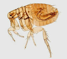 Fleas don’t just cause itching. They also carry infectious diseases that can be contagious to people. Controlling fleas on your pets protects your whole family’s health.
Tapeworms Tapeworms are parasites that live in the intestines. They shed small body segments called proglottids that pass out of the host animal’s body in the feces. Tapeworm segments in the stool look like whitish rice grains. Fleas transmit a type of tapeworm that commonly infects dogs and cats. Dogs and cats become infected by swallowing a flea. Tapeworms rarely cause significant disease in dogs and cats. The dog and cat tapeworm that is carried by fleas, Dipylidium caninum, can also infect humans (usually children) who swallow a flea.[1] Bartonellosis (Cat Scratch Disease) Bartonella species are bacteria that cause a variety of diseases in humans and other animals. Cat scratch disease and endocarditis (heart valve infection) are just 2 of the serious illnesses caused by Bartonella infection.[2] Fleas are the most common insect vector for Bartonella henselae, the species that causes cat scratch disease.[3] Fleas can also carry other Bartonella species. Infected cats and dogs might or might not have any symptoms of infection. Bartonellosis is a human health risk. Treating your cat with a cat-safe flea preventive reduces the risk of cat scratch disease for people in contact with your cat. Rickettsial Diseases Rickettsiae are a group of bacteria responsible for diseases such as typhus and Rocky Mountain spotted fever. Rickettsiae are spread by arthropods, including fleas and ticks. The types of fleas that infest dogs and cats transmit Rickettsia typhi (which causes murine typhus) and Rickettsia felis. Both of these bacteria can also cause disease in people.[1,4] Yersiniosis (Plague) Plague, including bubonic plague and the Black Death, is caused by infection with the bacterium Yersinia pestis. The Oriental rat flea (Xenopsylla cheopis) transmits the bacterium usually to rodents but sometimes to cats, dogs, other animals, and humans. Rat fleas in the western United States and other parts of the world still harbor Yersinia.[3] Mycoplasma Infection Cats infected with certain types of Mycoplasma bacteria develop anemia (low red blood cell count). Fleas are thought to be a source of infection for cats.[5] Noninfectious Diseases Fleas cause skin disease in animals that scratch or chew themselves to relieve the itch. Just a few fleas can set off intense itching in an animal with a flea allergy. Because fleas feed on blood, animals with lots of fleas can develop anemia from blood loss. This anemia can be life threatening. References 1. Fleas. Companion Animal Parasite Council website. https://capcvet.org/guidelines/fleas/. Updated September 19, 2017. Accessed May 7, 2019. 2. Bartonella infection (cat scratch disease, trench fever, and Carrión’s disease). Centers for Disease Control and Prevention website. https://www.cdc.gov/bartonella/index.html. Updated December 14, 2015. Accessed May 7, 2019. 3. Shaw SE. Flea-transmitted infections of cats and dogs. World Small Animal Veterinary Association World Congress Proceedings, 2008. Veterinary Information Network website. https://www.vin.com/doc/?id=3866578. Accessed May 7, 2019. 4. Little SE. Feline fleas and flea-borne disease (proceedings). DVM360 website. http://veterinarycalendar.dvm360.com/feline-fleas-and-flea-borne-disease-proceedings. Published April 1, 2010. Accessed May 7, 2019. 5. Lappin MR. Update on flea and tick associated diseases of cats. Vet Parasitol. 2018;254:26-29. Photo of Oriental rat flea (Xenopsylla cheopis) by James Gathany, CDC  We all know that cats sometimes throw up hairballs. But hairballs aren’t the only reason cats vomit. Don’t assume that vomiting is normal for your cat, even if it’s been going on for months or years.
Consult your veterinarian if your cat has any of these symptoms:
Hairballs Hairballs (trichobezoars in medical speak) are wads of hair in the digestive tract. Cats swallow loose hair when they lick their fur. Hair usually passes through the digestive tract without causing any problem. But sometimes hair packs together into a mass in the stomach or intestine. A hairball is usually shaped like a cylinder. If you see one on your favorite rug, you might mistake it at first for feces. Hairballs are often about the same size and shape as a log of cat poop. But if you look at a hairball closely you’ll see that it’s made of tightly packed hair (and it doesn’t smell like poop). We don’t really know how often cats normally vomit up hairballs. In an informal poll at a cat clinic in England, cat owners reported that nearly three-fourths of their cats had never vomited up a hairball. About 1 in 6 cats expelled a hairball once a year, and 1 in 10 cats expelled a hairball at least twice a year. Owners reported that long-haired cats brought up hairballs more frequently than short-haired cats did.[1] Hairballs that aren’t vomited up or passed in the stool can block the digestive tract. Symptoms include vomiting, decreased appetite, and signs of belly discomfort (which can be easy to miss in cats). Cats with intestinal blockages may need surgery. Causes of Hairballs Healthy cats occasionally bring up hairballs just because they’re cats and they groom themselves. But sometimes hairballs are a sign of another problem. Cats are more likely to have problems with hairballs if they swallow excessive amounts of hair or have a disorder that slows the movement of material through the digestive tract. Fleas and itchy skin conditions can lead to excessive grooming. Cats also overgroom in response to pain or stress. More grooming means more hair swallowed and an increased chance of hairballs. The symptoms of digestive tract diseases like inflammatory bowel disease and intestinal lymphoma (a type of cancer) can be mistaken for hairballs. In one study, some cats with hairballs blocking the intestines also had serious intestinal disease.[2] These diseases affect the way food and hair move through the digestive tract and may put cats at higher risk of hairballs. Hairballs can also irritate the digestive tract, causing inflammation. Managing Hairballs Ask your veterinarian before treating your cat’s vomiting or hacking with over-the-counter remedies or hairball diets. Hairballs might not be responsible for the symptoms. Brushing the coat to remove loose hair is safe and might be all that’s needed to reduce hairballs in healthy cats. Long-haired cats that regularly bring up hairballs may benefit from having their fur trimmed. If you’re noticing more hairballs than usual, ask your veterinarian to check your cat for underlying problems. If the symptoms point to an intestinal disease, your cat might need a series of diagnostic tests (such as bloodwork, ultrasound, and possibly a biopsy of the intestines). References 1. Cannon M. Hair balls in cats: a normal nuisance or a sign that something is wrong? J Feline Med Surg. 2013;15(1):21-29. 2. Norsworthy GD, Scot Estep J, Kiupel M, Olson JC, Gassler LN. Diagnosis of chronic small bowel disease in cats: 100 cases (2008-2012). J Am Vet Med Assoc. 2013;243(10):1455-1461. Photo by Karin Laurila |
AuthorLaurie Anne Walden, DVM Categories
All
Archives
April 2024
The contents of this blog are for information only and should not substitute for advice from a veterinarian who has examined the animal. All blog content is copyrighted by Mallard Creek Animal Hospital and may not be copied, reproduced, transmitted, or distributed without permission.
|
 RSS Feed
|
Office Hours
Monday through Friday 7:30 am to 6:00 pm
|
|


Site powered by Weebly. Managed by IDEXX Laboratories