Laurie Anne Walden, DVM  Dental radiography (x-ray imaging) is an important diagnostic tool for pets, just as it is for people. Animals don’t always show signs of mouth pain, and radiographs may be the only way to know there’s a problem. Most dental disease in dogs and cats happens under the gumline, where it’s hidden from view. In published studies, radiographs revealed disease in apparently healthy teeth in 27.8% of dogs and 41.7% of cats. Dental radiographs find problems that can’t always be seen on a physical examination:
Radiographs also guide dental procedures in pets. For example, some dog and cat teeth have curved roots, extra roots, or roots that extend very close to the jawbone. It’s best to know about these issues before trying to extract these teeth. In animals, dental radiography requires general anesthesia or sedation. The radiographs use sensors or small film cassettes placed inside the mouth, and animals can’t wiggle during the procedure. Imagine putting an x-ray sensor between your dog’s teeth and politely asking her to lie still and not chew on it. Now imagine asking your cat the same thing. Dental radiographs are often obtained while a pet is already under anesthesia for a dental procedure. Dental radiography delivers a very small radiation dose to the patient. The American Veterinary Dental College reports that “the radiation risk to the patient from taking dental radiographs is minimal.” Like people, pets benefit from having full-mouth dental radiography from time to time to be sure no problems are lurking under the surface. Oral imaging may also be indicated in pets with these conditions:
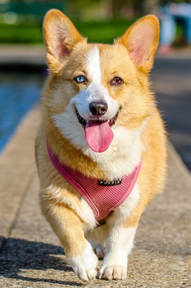
Photo by muhannad alatawi Comments are closed.
|
AuthorLaurie Anne Walden, DVM Categories
All
Archives
June 2024
The contents of this blog are for information only and should not substitute for advice from a veterinarian who has examined the animal. All blog content is copyrighted by Mallard Creek Animal Hospital and may not be copied, reproduced, transmitted, or distributed without permission.
|
- Home
- About
- Our Services
- Our Team
-
Client Education Center
- AKC: Spaying and Neutering your Puppy
- Animal Poison Control
- ASPCA Poisonous Plants
- AVMA: Spaying and Neutering your pet
- Biting Puppies
- Boarding Your Dog
- Caring for the Senior Cat
- Cats and Claws
- FDA warning - Bone treats
- Force Free Alliance of Charlotte Trainers
- Getting your Cat to the Vet - AAFP
- Holiday Hazards
- How To Feed Cats for Good Health
- How to Get the Most Out of your Annual Exam
- Indoor Cat Initiative - OSU
- Introducing Your Dog to Your Baby
- Moving Your Cat to a New Home
- Muzzle Training
- Osteoarthritis Checklist for Cats
- What To Do When You Find a Stray
- Our Online Store
- Dr. Walden's Blog
- Client Center
- Contact
- Cat Enrichment Month 2024
|
Office Hours
Monday through Friday 7:30 am to 6:00 pm
|
Mallard Creek Animal Hospital
2110 Ben Craig Dr. Suite 100
|
Site powered by Weebly. Managed by IDEXX Laboratories

 RSS Feed