Laurie Anne Walden, DVM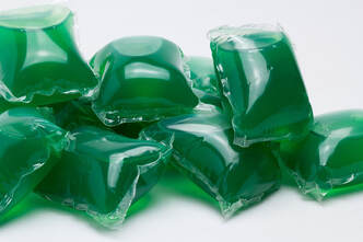 Photo by Antonio Jose Cespedes on Pixabay Photo by Antonio Jose Cespedes on Pixabay Laundry and dish detergents contain chemicals that are unsafe for animals. Detergent pods pose a bigger risk than bottled liquid detergents or detergent powders.
Toxic Ingredients Detergents contain anionic and nonionic surfactants, which lift dirt and oil off of surfaces and fabrics. These types of surfactants cause mild irritation to the skin and eyes. If swallowed, they can cause vomiting. Fabric softeners, disinfectants, and sanitizing solutions sometimes contain cationic surfactants, which are more corrosive than anionic and nonionic surfactants. Cats are especially sensitive to cationic surfactants. Direct contact can injure the skin, and cats that lick these products from their paws or fur can also have damage to the mouth and digestive tract. Some detergents are alkaline (they have a high pH, the opposite of acids). Concentrated alkaline products damage the stomach if swallowed. Detergents might also contain ethanol and other potentially toxic ingredients. Detergent Pods Animals (mainly dogs) that bite detergent pods are at higher risk than animals that lick liquid detergents or detergent powders. Even when the ingredients are similar, the consequences of exposure are more serious with detergent packaged in pods. Detergent in pods is concentrated and under pressure. When a tooth punctures a pod, the detergent sprays the inside of the mouth, from where it is swallowed, inhaled into the lungs, or both. Animals that lick spilled liquid detergent aren’t likely to consume very much of it because it doesn’t taste good. In comparison, dogs that bite detergent pods get a larger dose of product at a higher concentration. The most severe effects of detergent exposure are lung inflammation and pneumonia, which happen when detergent—or vomit that contains detergent—is inhaled into the lungs. Pneumonia can be fatal, so it’s possible for dogs that chew detergent pods to die. Signs of Detergent Toxicosis
If Your Pet Is Exposed
Sources
Image source: Antonio Jose Cespedes on Pixabay Laurie Anne Walden, DVM Photo by Wall Boat on Flickr Photo by Wall Boat on Flickr Uveitis is inflammation inside the eye. Some of the many conditions that cause uveitis affect the whole body, so a diagnosis of uveitis often triggers a series of tests to find the cause.
The uvea is a pigmented layer within the eye. The iris, which gives the eye its color, is the part of the uvea that you can see when you look at your pet’s eyes. The other structures of the uvea (ciliary body and choroid) are behind the iris, sandwiched between other layers of the eye. The uvea has a blood supply to deliver nutrients to the eye, so anything that can travel through blood vessels—like infectious organisms and cancer cells—can reach the uvea and cause uveitis. The uvea also produces and drains fluid within the eye. Inflammation of the uvea interferes with fluid balance inside the eye, so untreated uveitis can lead to glaucoma (increased eye pressure) and blindness. Causes Some of the things that cause uveitis are problems with the eye itself:
Problems outside the eye can also cause uveitis:
Signs Uveitis causes the same nonspecific signs of eye trouble (like redness) as nearly every other eye disorder. It can also cause changes that are more specific to inflammation inside the eye, like a change in iris color. Animals with uveitis sometimes have no obvious signs at all. These are some of the signs of uveitis:
Diagnosis Uveitis is diagnosed by examining the eye with an ophthalmoscope to look for signs of inflammatory debris inside the eye. The eye exam might be the only diagnostic test needed if it reveals an eye problem that’s causing the uveitis. If the animal doesn’t have an obvious eye abnormality causing the inflammation, the next step is to conduct diagnostic tests to find the cause. The animal will most likely have baseline blood tests, tests for various infectious organisms (especially viruses and the many organisms carried by fleas and ticks), and possibly x-ray imaging or ultrasound. The patient might need to be referred to a veterinary ophthalmologist. Treatment Uveitis is treated with medication to reduce inflammation. This medication is usually in the form of eye drops or eye ointment. Some animals need oral medication in addition to eye medication. The purpose of this anti-inflammatory medication is to relieve pain and reduce the risk of glaucoma. Whatever caused the uveitis also needs to be treated. The prognosis for uveitis depends on the cause. If uveitis is diagnosed quickly and the underlying cause is treatable, the prognosis is generally good. Some conditions that cause uveitis have a more guarded prognosis. Image source: Wall Boat on Flickr Laurie Anne Walden, DVM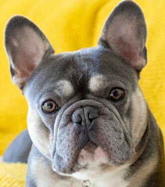 This French bulldog has narrowed nostrils and prominent facial skin folds. Photo by Dan Blackburn on Unsplash. This French bulldog has narrowed nostrils and prominent facial skin folds. Photo by Dan Blackburn on Unsplash. The French bulldog is now the most popular dog in the United States, according to the American Kennel Club (which defines popularity by the number of purebred dog registrations). This result is no surprise; Frenchies have been rising in the popularity ranks for several years. But these little dogs have a host of potential health problems, just like other animals bred to have flat faces and short noses.
Brachycephalic Head Shape Short-headed (brachycephalic) animals like English bulldogs, French bulldogs, pugs, and Persian cats have a shortened upper jaw and nose. The lower jaw is typically not shortened, so when they close their mouths, their lower incisor teeth might stick out in front of the upper incisors. Brachycephaly affects only the bones, not the soft tissues (skin, tongue, soft palate, and so forth), so brachycephalic animals have too much soft tissue for their face size. This is why brachycephalic animals have skin folds between their nose and eyes. They have the skin to cover a nose that just isn’t there. Respiratory Problems In a brachycephalic animal, the soft tissues inside the mouth and throat are crammed into an upper jaw that’s too short to hold them. All of this excess tissue blocks the airway, causing a cascade of problems related to the increased effort of breathing. The term brachycephalic airway syndrome describes problems caused by anatomic abnormalities that are common in brachycephalic animals. These abnormalities include narrowed nostrils, an elongated soft palate, and everted laryngeal saccules (tissue near the vocal cords that is pulled into the airway because of labored breathing over time). Brachycephalic animals might also have an enlarged tongue. Some have an abnormally narrow trachea, which increases the work of breathing and the risk of problems during anesthesia. Other Problems Brachycephalic animals are more likely than others to have heat stress. Their relatively shallow eye sockets and large eyelid openings increase their chance of developing corneal ulcers, dry eye, and other eye problems. Infections can develop between skin folds. Some brachycephalic animals have digestive tract problems like chronic vomiting, possibly related to gastric reflux caused by chronic labored breathing. They usually have crowded or malpositioned teeth. Natural birth is not possible for some brachycephalic dogs because the mom’s pelvis is too narrow for the puppies’ heads. Some breeds—including French bulldogs—usually require cesarean (surgical) delivery. Signs If you have a brachycephalic pet, watch for these signs of trouble:
Treatment Anatomic problems like narrowed nostrils, elongated soft palate, and everted laryngeal saccules can be corrected with surgery. You can see photos of these problems and more information about surgery on the American College of Veterinary Surgeons website: https://www.acvs.org/small-animal/brachycephalic-syndrome. One “treatment” for brachycephalic airway syndrome is to stop breeding dogs for extreme face shape. If you’re thinking of getting a French bulldog or another brachycephalic pet, reward breeders who breed for good health: choose a dog with round (not slit-like) nostrils, minimal or no skin folds near the eyes, and the ability to run, play, and sleep while breathing freely without snorting or gagging. Image source: Dan Blackburn on Unsplash Laurie Anne Walden, DVM Photo by Hermes Rivera on Unsplash Photo by Hermes Rivera on Unsplash Eyelid problems are common in dogs and cats. Some eyelid disorders cause damage to the eyeball itself and can affect vision. A pet with eye redness, eye drainage, eyelid swelling, an eyelid lump that is new or changing, or signs of eye discomfort (squinting, pawing at the eyes, rubbing the face) should see a veterinarian.
Benign Masses In dogs, most eyelid masses are benign tear gland tumors called meibomian gland adenomas. These masses are located along the lid margin and typically look like raised, bumpy, pink or dark brown lumps. Other types of benign tumors also occur in dogs and cats. A chalazion is a firm lump caused by a blocked tear gland. This type of lump is near the lid margin under the skin, not right on the lid margin like a meibomian gland adenoma. Benign eyelid masses can irritate the cornea (the clear structure at the front of the eye) and can enlarge over time. These masses are usually removed surgically. Removing an eyelid mass means removing part of the eyelid, so it’s best to do the procedure while the mass is still small. Cancerous Masses Most eyelid masses in cats, and some in dogs, are malignant cancers. Many types of cancer affect the eyelids, just like the rest of the skin; some examples are squamous cell carcinoma, melanoma, and mast cell tumor. The only way to know for sure if an eyelid lump is benign or malignant is to send it to a laboratory for analysis. If an eyelid mass is large or located near the inner corner of the eye, referral to a veterinary ophthalmologist is often the best option in case the patient needs reconstructive eyelid surgery. Cherry Eye The term cherry eye is an informal name for prolapse of the gland of the third eyelid. The third eyelid (nictitating membrane) is a pink structure at the inner corner of dogs’ and cats’ eyes. In animals with cherry eye, a tear gland protrudes out of its normal location behind the third eyelid; it looks like a smooth pink or red lump. Cherry eye is treated by surgically replacing the gland in its normal position. The prolapsed gland shouldn’t be removed entirely because it produces tears. Removal of the gland would put the animal at risk of dry eye (keratoconjunctivitis sicca). Swollen Eyelids Eyelid swelling could be caused by an allergic reaction, a benign or cancerous mass, inflammation, or infection. Eyelid swelling due to an allergic reaction is sudden and often affects both eyes. Because eyelid swelling is sometimes the first sign of a more serious reaction called anaphylaxis, it always warrants a call or visit to a veterinary clinic. An animal that has swollen eyelids along with other concerning symptoms like vomiting or wheezing should be taken immediately to an emergency clinic. Entropion (Rolled-in Eyelids) In animals with entropion, one or more eyelids are rolled inward so eyelashes and hair rub on the cornea. Entropion can be genetic or breed associated, especially in animals with loose facial skin and animals that are brachycephalic (short faced). The condition can also be caused by scarring, other eyelid disorders, or excessive squinting from eye discomfort. The damage to the cornea is uncomfortable at best and can eventually impair vision. Entropion is corrected with surgery. Young animals with entropion can sometimes be treated with a simple tacking procedure to help the eyelids roll back out to the normal position as they grow. Abnormal Hairs Occasionally hairs similar to eyelashes grow in the wrong location, either along the eyelid margin (distichia) or inside the eyelid (ectopic cilia). These hairs can cause significant pain and corneal ulcers. They’re hard to see and are typically found only with magnification during an ophthalmic examination to find out why an animal has a corneal ulcer. If they’re causing discomfort, the hairs and their follicles need to be removed. Eyelid Inflammation Inflamed eyelids are red and swollen, sometimes with oozy discharge and visible sores along the margins. The hair on the eyelid might be thin or missing. Eyelid inflammation has many possible causes, including immune-mediated disease, parasites (like mange mites), bacterial or fungal infections, and irritants. Diagnosis might require skin scrapings, cultures, and biopsy. Treatment depends on the underlying cause. More Information You can see photos of some of these conditions on the American College of Veterinary Ophthalmologists website: https://www.acvo.org/common-conditions1 Image source: Hermes Rivera on Unsplash Laurie Anne Walden, DVM Photo by Jennifer Shishmanian on Unsplash Photo by Jennifer Shishmanian on Unsplash Keratoconjunctivitis sicca (KCS), or dry eye, is common in dogs and can also affect cats. KCS is caused by a problem with the quantity or quality of the tear film. The condition is uncomfortable and can cause blindness. For most patients, KCS is managed with lifelong eye medication.
Causes Tears are produced by glands in the eyelids and third eyelid. The tear film contains water, mucus, and oil. Deficiencies in any of the components of the tear film compromise its functions: lubricating the cornea (the clear structure at the front of the eye), removing debris, and providing nutrition to the cornea. Anything that affects the tear-producing glands can cause KCS. Brachycephalic (flat-faced) animals with protruding eyes that aren’t completely covered by the eyelids also develop KCS symptoms because of excessive water evaporation from the surface of the cornea. These are some of the causes of KCS:
Signs Animals with KCS typically have thick, sticky eye discharge. The discharge is thick because the watery part of the tear film is decreased or absent, so the tear film that remains is made mostly of mucus and oil. Either 1 or both eyes are affected, depending on the cause of the KCS. Tear film deficiencies increase the risk for corneal ulcers, long-term damage to the cornea, and impaired healing of the cornea. These are signs of KCS and the resulting corneal damage:
Diagnosis The tear film quantity is measured with the Schirmer tear test, in which a sterile paper strip with measurement markings is placed inside the lower eyelid for 1 minute. The distance that the tear film is absorbed along the strip shows whether the animal has low tear production. Veterinary ophthalmologists use other tests to find out whether a patient has a problem with tear film quality. Treatment Animals with KCS are treated with a combination of eye medications, most of which need to be applied 2 or 3 times a day. Medications include tear stimulants that reduce immune-mediated inflammation, tear replacements to lubricate the eye, and other medications to treat corneal damage. KCS can be managed but not cured, so medical treatment needs to continue for the rest of the animal’s life. For animals whose KCS doesn’t respond to eye medication, a few surgical options are available. The most common of these is parotid duct transposition, which involves rerouting a salivary duct from the mouth to the inside of the lower eyelid so the animal basically drools onto the eye. This procedure is performed by a veterinary ophthalmologist. Image source: Jennifer Shishmanian on Unsplash Laurie Anne Walden, DVM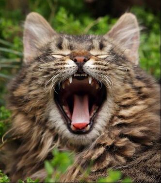 Photo by mahyar mirghasemi Photo by mahyar mirghasemi Dogs and cats need regular dental care, including thorough oral exams and dental cleanings. These procedures are best done under general anesthesia, but some pets don’t receive the dental care they need because their owners are worried about the safety of anesthesia. Most animals handle anesthesia well, though, and the risk is greatly reduced with preanesthetic testing, safe anesthetic drugs, and monitoring during the procedure.
Reasons for Dental Anesthesia General anesthesia during a dental procedure allows for better medical care, reduces an animal’s stress and pain, and makes the procedure safer for everyone.
Anesthesia Safety Before an animal undergoes anesthesia, the benefits of the procedure are weighed against the possible risks. Although general anesthesia is safe overall, its risks range from minor drug effects that can be anticipated and managed to potentially fatal complications, which are rare. Animals receive a thorough physical exam before anesthesia and might also have preanesthetic laboratory tests. Preanesthetic tests are especially important for senior animals—the age group most often having dental procedures—because liver, kidney, and heart function all affect anesthesia risk. During the procedure, vital signs like heart rate, respiratory rate, blood oxygen saturation, and blood pressure are monitored. The patient’s level of anesthesia is also monitored and can be adjusted as needed. If your pet will be having anesthesia, tell the veterinarian all of the medications and supplements your pet is getting at home. Your veterinarian will also need to know if your pet has ever had problems with anesthesia in the past. If your pet has been to more than one clinic, be sure the anesthetizing veterinarian has your pet’s complete medical records and knows of any history of breathing trouble, heart conditions, seizures, or other medical problems. Problems With Anesthesia-Free Dental Scaling Some places offer dental scaling without anesthesia, but this procedure isn’t in an animal’s best interest.
Photo by mahyar mirghasemi on Unsplash Laurie Anne Walden, DVM Polyurethane glues that expand when they’re exposed to moisture pose a serious risk to dogs that chew the container and swallow even a little bit of the product. Some (not all) Gorilla Glue products are expanding adhesives. Some wood glues, craft glues, general household glues, and construction adhesives also expand when wet.
Dogs rarely swallow expanding adhesives, but the potential consequences can be severe. Anything that swells up when it’s wet is hazardous if swallowed. With expanding adhesives, the risk is even greater: these products expand to a size much larger than the amount swallowed and then harden to form a solid mass that can block material from entering or leaving the stomach. Expanding adhesives contain diisocyanates, which are chemicals used to make polyurethane products. Diisocyanates are hygroscopic, meaning they absorb moisture from the air. Glues that contain diisocyanates are used to seal cracks and fill gaps. In the moist, acidic environment of the stomach, products containing diisocyanates expand up to 8 times their original volume. Expansion starts within minutes after the product is swallowed. The hardened mass of glue becomes a foreign object in the stomach. A volume of glue as small as half an ounce can form a mass large enough to cause a blockage.[1] Glue that has cured before being swallowed—has already finished expanding and has formed a solid mass—is much less of a chemical risk, although of course it could still cause an obstruction if a dog managed to swallow a big enough chunk. Diisocyanates also irritate the skin and respiratory system, but these problems are much less common in animals than in people who are exposed to high levels at work. Signs and Diagnosis A mass of glue in the stomach causes the same signs as any other foreign object: vomiting, loss of appetite, painful abdomen, and loss of energy. Glue ingestion is diagnosed, or at least strongly suspected, when a dog is seen swallowing glue, the dog’s owner finds a chewed glue container, or the dog has glue residue on the fur. The mass formed by a diisocyanate glue is visible on radiographs (x-ray images) but can look very similar to food in the stomach. Other imaging tests or a series of radiographs taken over time might be needed to definitely diagnose a foreign object in the stomach. Expanding adhesives don’t stick to the inside of the stomach, luckily. In a study of dogs treated for Gorilla Glue ingestion, the glue irritated the stomach lining and caused ulcers in some dogs but didn’t cause serious damage to the stomach in any dog. The dogs had bloodwork abnormalities similar to those caused by vomiting in general or any type of stomach obstruction. Whether the presence of diisocyanates in the stomach caused any other problems wasn’t clear.[1] Treatment A foreign object that’s causing a stomach obstruction needs to be surgically removed. If a mass of glue seems small enough to pass on its own and the dog doesn’t have any worrisome signs like belly pain, the veterinarian and dog’s owner might decide to watch and wait. What to Do if Your Pet Is Exposed
Reference 1. Friday S, Murphy C, Lopez D, Mayhew P, Holt D. Gorilla Glue ingestion in dogs: 22 cases (2005-2019). J Am Anim Hosp Assoc. 2021;57(3):121-127. doi:10.5326/JAAHA-MS-7126 Laurie Anne Walden, DVM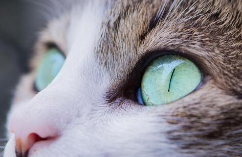 Photo by Antonio Lapa on Unsplash Photo by Antonio Lapa on Unsplash The conjunctiva is a thin membrane that covers the insides of the eyelids and the white part of the eye. Inflammation of the conjunctiva, or conjunctivitis, makes the eye look red. Conjunctivitis is sometimes called pinkeye. This name is a bit misleading, though, because lots of other eye disorders—some very serious—also cause redness of the eye.
The conjunctiva is a mucous membrane, like the membranes lining the nose and mouth. It protects the eye, helps lubricate the surface of the eye with tears, aids in eye movement, and is part of the system that heals damage to the cornea. Causes Conjunctivitis has many possible causes in dogs and cats:
Signs Signs include eye redness, squinting, blinking more than usual, and eye discharge. The discharge can be watery and clear or it can be cloudy, yellow, or greenish. The tissues around the white of the eye and the insides of the lids might look puffy. The animal might rub or paw at the eye because of itching or discomfort. Diagnosis Conjunctivitis can’t be diagnosed just by the appearance of the eye. Redness and discharge are part of the eye’s response to any abnormality, so other problems need to be ruled out before conjunctivitis can be diagnosed. Diagnosis includes a general physical examination, an eye examination with an ophthalmoscope, and a corneal stain to identify ulcers or other damage to the cornea. Other tests include measuring tear production, measuring eye pressure (to rule out glaucoma), and sometimes swabbing or scraping the conjunctiva to obtain samples for laboratory analysis. Treatment Conjunctivitis is treated with topical medication (eye drops or eye ointment); oral medication is sometimes also used. The type of medication depends on the cause of the conjunctivitis. Antibiotics are usually part of the treatment, either to eliminate a primary bacterial infection or to prevent a secondary bacterial infection. Cats with known or suspected herpesvirus conjunctivitis sometimes need antiviral medication. Eye medications used to treat conjunctivitis often contain a steroid (such as dexamethasone or hydrocortisone) to reduce inflammation. However, steroids interfere with corneal healing, so before a steroid-containing medication is used, the cornea must be stained and examined with an ophthalmoscope to be sure there is no corneal ulcer or scratch. If your pet has eye medication from an earlier episode of conjunctivitis, don’t use it to treat a new episode of eye redness unless your veterinarian has instructed you to. And never use any over-the-counter (nonprescription) eye product in an animal without first consulting your veterinarian. Photo by Antonio Lapa on Unsplash Laurie Anne Walden, DVM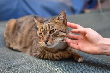 Photo by Ilze on Unsplash Photo by Ilze on Unsplash Aggression related to being petted is very common in cats. Cats with this type of aggression suddenly scratch, growl, or bite while they’re being petted.
The most likely explanations for petting-related aggression are that the cat is overstimulated, has a low tolerance for being touched, or is trying to exert some control over the interaction. It’s also possible that the cat has a medical problem (for example, pain caused by arthritis or dental disease). Any type of aggression, especially if it’s a new behavior, warrants an examination by a veterinarian. The people most at risk of injury are children and others who don’t know the signs that a cat is uncomfortable with an interaction. Tolerance to petting varies from cat to cat, and the same cat’s tolerance can change according to the circumstances. To help people understand the best way to interact with cats, a team of researchers in the United Kingdom developed a set of guidelines that they describe with the acronym CAT (for choice, attention, and touch).[1] The team found that cats showed less aggression when people followed the CAT guidelines during interactions. The gist of the CAT guidelines is that we should handle cats the way they want to be handled and leave them alone when they want to be left alone. Makes sense, right? However, in another study, the same team found that some people who had lots of cat-owning experience and rated their own cat knowledge as high interacted in ways the cats did not want.[2] The following is a summary of the CAT guidelines, or how to pet a cat. C: Give the cat choice and control.
A: Pay attention to the cat’s body language. These are signs that a cat might not want to be petted any longer:
T: Touch the cat only where the cat wants to be touched.
To read more about cat-friendly petting and how humans interact with cats, check out these 2 articles:
References 1. Haywood C, Ripari L, Puzzo J, Foreman-Worsley R, Finka LR. Providing humans with practical, best practice handling guidelines during human-cat interactions increases cats' affiliative behaviour and reduces aggression and signs of conflict. Front Vet Sci. 2021;8:714143. doi:10.3389/fvets.2021.714143 2. Finka LR, Ripari L, Quinlan L, et al. Investigation of humans individual differences as predictors of their animal interaction styles, focused on the domestic cat. Sci Rep. 2022;12(1):12128. doi:10.1038/s41598-022-15194-7 Photo by Ilze on Unsplash Laurie Anne Walden, DVM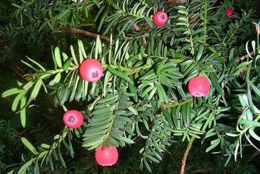 Yew is highly toxic to animals and people. Yew is highly toxic to animals and people. Some decorative winter holiday plants pose risks to pets. Other plants that aren’t actually toxic can still cause upset stomach if an animal swallows them.
If you think your pet might have swallowed or been exposed to a toxic plant, contact your veterinarian, an animal emergency clinic, or a 24-hour animal poison control hotline (a fee may apply):
Amaryllis (Amaryllis species) Ingestion of amaryllis leaves, stems, or bulbs can cause drooling, vomiting, diarrhea, belly pain, lethargy, and a drop in blood pressure. Christmas cactus (Schlumbergera species) Christmas cactus isn’t toxic. However, an animal that swallows part of one might vomit or have diarrhea. Christmas rose, hellebore (Helleborus niger) Hellebores contain cardiac glycosides, compounds that affect heart function. Ingestion of any part of the plant can cause drooling, belly pain, vomiting, diarrhea, and lethargy. Christmas trees The Christmas trees most commonly grown in North Carolina (firs, pines, cedars, and cypress) aren’t toxic, although like other non-food plants they could cause vomiting or intestinal blockage if an animal swallowed enough of one. The risks to pets are from trees falling, ornaments breaking, exposure to electrical cords, and possibly exposure to preservatives (which are not very toxic but can cause mild vomiting, according to ASPCA Animal Poison Control). Norfolk Island pines and rosemary plants are often sold as potted miniature holiday trees. These two plants aren’t toxic to cats and dogs. However, a miniature tree made of mixed greenery would be a serious danger if it contained yew. Cyclamen (Cyclamen species) Ingestion of cyclamen, a flowering houseplant, can cause drooling, vomiting, and diarrhea. Swallowing a large amount of cyclamen tubers can lead to heart rhythm abnormalities, seizures, and death. Delphinium, larkspur (Delphinium species) Blue delphinium flowers are included in some Hanukkah floral arrangements. Toxic compounds in delphinium block a neurotransmitter that’s required for muscle function. Ingestion can cause digestive system problems (vomiting, diarrhea, or constipation), belly pain, drooling, weakness, abnormal heart rhythm, tremors, seizures, and paralysis. The most severe effects—heart or lung failure and death—are most likely to happen in grazing animals that ingest large amounts of the plant. Holly, winterberry, Christmas holly, English holly (Ilex aquifolium) Ingestion of English holly and similar plants in the genus Ilex can cause vomiting and diarrhea. The pointed leaves can injure the inside of the mouth, leading to drooling and other signs of mouth discomfort. Lily (Lilium species, Hemerocallis species) Lilies of various types are common in floral arrangements, including arrangements for winter holidays. Some lilies are so toxic to cats that they shouldn’t be brought at all into a house with cats. The most dangerous lilies are Lilium species (Easter lily, Japanese lily, Asiatic hybrid lilies, stargazer lily, Casablanca lily, and tiger lily) and Hemerocallis species (daylily). Ingestion of a tiny amount of plant material—even licking pollen from a paw—can cause kidney injury in cats. Calla lilies, peace lilies, lily of the valley, and Peruvian lilies are not Lilium and Hemerocallis species; these plants don’t damage the kidneys but can cause stomach upset and other problems. Mistletoe (Phoradendron species, Viscum album) Mistletoe ingestion can cause vomiting, diarrhea, and abdominal pain in animals. Swallowing large amounts can cause low heart rate, a drop in blood pressure, uncoordinated gait, and seizures. American mistletoe (Phoradendron species) is less toxic than European mistletoe (Viscum album). Paperwhite (Narcissus papyraceus) Paperwhites and other Narcissus species (like jonquils and daffodils) cause severe vomiting and diarrhea if ingested. Swallowing a large amount can lead to breathing and heart rhythm abnormalities. The most toxic part of the plant is the bulb. Poinsettia (Euphorbia pulcherrima) Poinsettias are not as toxic as some have been led to believe, according to ASPCA Animal Poison Control. The thick, milky sap is an irritant that can cause drooling and vomiting if ingested. The sap can also irritate the skin and eyes on contact. Yew (Taxus species) Plants in the genus Taxus are highly toxic to all animals, including dogs, cats, horses, and people. Yew branches and berries are sometimes used to make holiday wreaths and other decorations. Be very careful displaying and disposing of items that might contain yew; be sure pets and wildlife can’t access them. Yew ingestion causes vomiting, muscle tremors, difficulty breathing, heart failure, seizures, and death. Sources
Public domain photo of yew (Taxus baccata) by MM on Wikimedia Commons |
AuthorLaurie Anne Walden, DVM Categories
All
Archives
June 2024
The contents of this blog are for information only and should not substitute for advice from a veterinarian who has examined the animal. All blog content is copyrighted by Mallard Creek Animal Hospital and may not be copied, reproduced, transmitted, or distributed without permission.
|
- Home
- About
- Our Services
- Our Team
-
Client Education Center
- AKC: Spaying and Neutering your Puppy
- Animal Poison Control
- ASPCA Poisonous Plants
- AVMA: Spaying and Neutering your pet
- Biting Puppies
- Boarding Your Dog
- Caring for the Senior Cat
- Cats and Claws
- FDA warning - Bone treats
- Force Free Alliance of Charlotte Trainers
- Getting your Cat to the Vet - AAFP
- Holiday Hazards
- How To Feed Cats for Good Health
- How to Get the Most Out of your Annual Exam
- Indoor Cat Initiative - OSU
- Introducing Your Dog to Your Baby
- Moving Your Cat to a New Home
- Muzzle Training
- Osteoarthritis Checklist for Cats
- What To Do When You Find a Stray
- Our Online Store
- Dr. Walden's Blog
- Client Center
- Contact
- Cat Enrichment Month 2024
 RSS Feed
|
Office Hours
Monday through Friday 7:30 am to 6:00 pm
|
Mallard Creek Animal Hospital
2110 Ben Craig Dr. Suite 100
|
Site powered by Weebly. Managed by IDEXX Laboratories